HIV infections are particularly difficult to clear for many reasons, one being that they hide out in the immune system’s own cells. In fact, a specific type of cells called follicular helper T cells (Tfh cells) appear to be a particularly fertile “reservoir” for HIV, and scientists are interested in empowering these cells to better fight off HIV.
There are two main types of T cells in the immune system, helper cells and antiviral, killer cells that differ based on the molecules they express on their cell surface and the part they play in the immune system. Normally antiviral T cells do not have access to germinal centers where HIV can persist in an HIV-positive person despite their taking antiretroviral drugs.
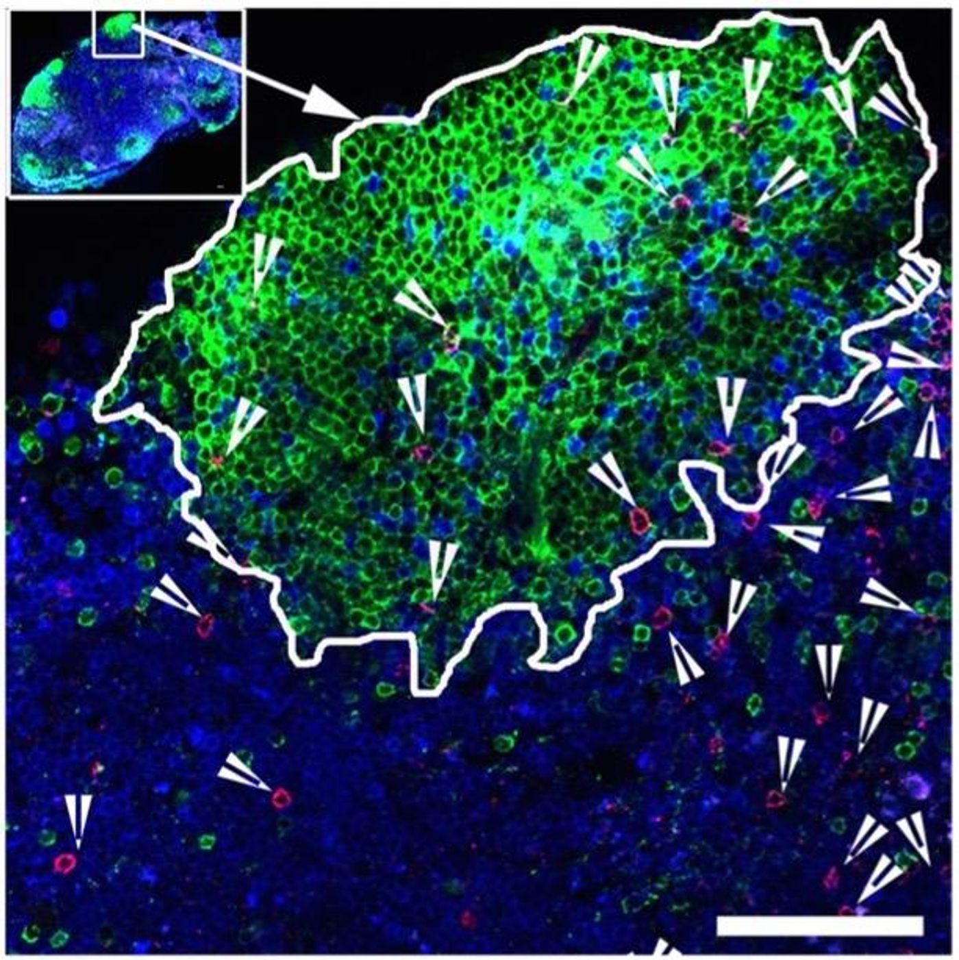
However, a new study from the Yerkes National Primate Research Center at Emory University identified a group of antiviral T cells that are essentially given “badge access” into germinal centers, thanks to a molecule expressed on their cell surface called CXCR5. And if CXCR5 is the key to T cell access to germinal centers, is there a way to induce CXCR5 expression on the surface of antiviral T cells? This was the goal of researcher Rama Rao Amara, PhD, with the idea that CXCR5 display would boost the anti-HIV ability of the immune system.
"These cells have the potential to infiltrate sites of ongoing viral replication and persistence," Amara said of Tfh cells, which express CXCR5 on their surface and have access to germinal centers. "Their properties and the ability to induce these cells by vaccination provide a tremendous opportunity to target and reduce the viral reservoir in lymphoid tissues."
Amara and a team of researchers from Emory conducted a study with rhesus macaques, which were vaccinated against SIV, the “monkey version” of HIV, followed by consistent exposure to the virus. While a promising proportion of macaques showed nearly 40 percent of CXCR5-expressing antiviral T cells in their lymph nodes, some of the animals ended up with varying degrees of infection despite being vaccinated against SIV.
From the study, researchers concluded that the more CXCR5-positive antiviral T cells, the better the immune system’s ability to control the virus, and Amara calls antiviral T cells displaying CXCR5 “functionally superior.” And although sometimes when stimulated, CXCR5-positive cells lose their CXCR5 expression, an immune regulatory molecule called TGF-beta can fix this problem.
They also noticed that the Tfh cells that have natural access to germinal centers by displaying CXCR5 have stem cell-like properties, explained co-corresponding author Vijayakumar Velu, PhD, because they can differentiate into both CXCR5-positive and -negative cells. Interestingly, the same type of cells appear to have been found in mice with other chronic viral infections.
The recent study was published in the journal PNAS.
Source: Emory Health Sciences