Immune cells in the uterus that produce important growth-promoting factors could be both the explanation for and the solution to recurrent spontaneous abortions affecting pregnant women worldwide. In a new study published in the journal Immunity, scientists explore natural killer cells in the uterus.
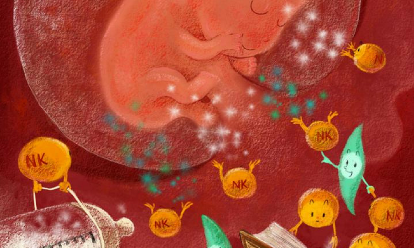
Natural killer (NK) cells are some of the most plentiful cells in the uterus during pregnancy, but only during the first trimester; the NK cell population decreases after formation of the placenta. However, while NK cells remain abundant, a specific subset of these cells plays the vital role of producing pleiotrophin and osteoglycin, growth-promoting factors found in both humans and mice.
Co-senior author Haimin Wei praises the study for both identifying new characteristics of natural killer cells in the context of pregnancy and for highlighting “approaches for therapeutic administration of natural killer cells in order to reverse restricted nourishment within the uterine microenvironment.”
Pregnant women with a smaller-than-normal population of pleiotrophin- and osteoglycin-producing NK cells in the uterine lining were found to be more likely to experience recurrent spontaneous abortion, also known as recurrent miscarriage, habitual abortion, or recurrent pregnancy loss; it occurs in about 15 percent of all “clinically recognized” pregnancies. This condition is thought to happen due to what Wei calls “restricted” fetal development as a result of missing growth-promoting factors from NK cells.
Wei and other researchers also found that supplying pregnant mice with these NK cells could reverse damage done to fetal development as a result of deficient amounts of pleiotrophin and osteoglycin. As a clinical application, medical professionals could potentially transfer natural killer cells via intravenous infusion or the administration of a vaginal suppository, Wei says - no need for invasive procedures with pregnant women.
Going forward, Wei and his team have yet to determine whether pleiotrophin, osteoglycin, and other growth-promoting factors impact fetal development directly, via the maternal-fetal barrier, or indirectly, via support of placenta and blood vessel growth.
"This study provides an avenue for treating fetal growth restriction, recurrent spontaneous abortion with unknown reasons, and age-related fetal loss by improving the uterus microenvironment,” concluded co-senior author Zhigang Tian.
Sources: Reviews in Obstetrics & Gynecology, Cell Press