A team of researchers announced in the journal
Science that they’ve determined the structure of the Zika virus and found regions where it differs from similar viruses such as West Nile and dengue.
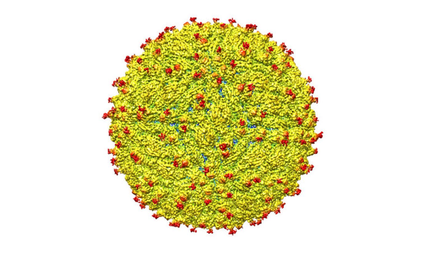
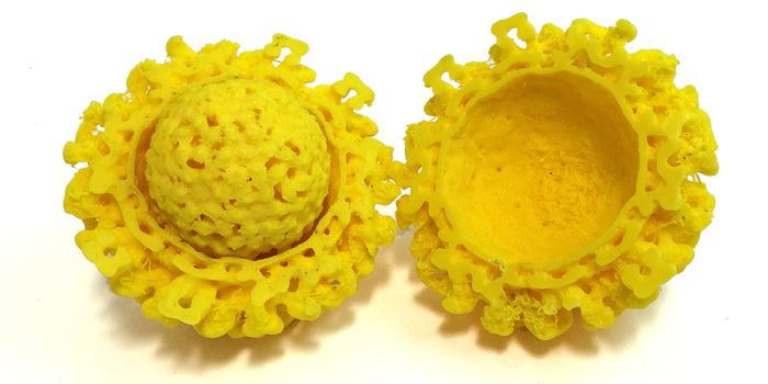
Image Credit: Kuhn and Rossmann research groups/Purdue University
Those differences could potentially explain how the virus is transmitted and how it causes disease, according to Richard Kuhn, who co-led the team and directs the Purdue Institute for Inflammation, Immunology, and Infectious Diseases.
“The structure of the virus provides a map that shows potential regions of the virus that could be targeted by a therapeutic treatment, used to create an effective vaccine or to improve our ability to diagnose and distinguish Zika infection from that of other related viruses,” says Kuhn. “Determining the structure greatly advances our understanding of Zika—a virus about which little is known. It illuminates the most promising areas for further testing and research to combat infection.”
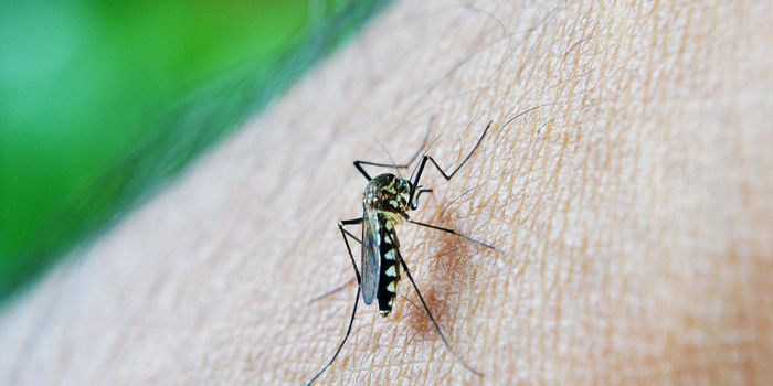
The Zika virus, a mosquito-borne disease, has recently been associated with a birth defect called microcephaly that causes brain damage and an abnormally small head in babies born to mothers infected during pregnancy. It also has been associated with the autoimmune disease Guillain-Barré syndrome, which can lead to temporary paralysis.
In the majority of infected individuals symptoms are mild and include fever, skin rashes and flulike illness, according to the World Health Organization.
Zika virus transmission has been reported in 33 countries. Of the countries where Zika virus is circulating, 12 have reported an increased incidence of Guillain-Barré syndrome, and Brazil and French Polynesia have reported an increase in microcephaly, according to WHO. In February WHO declared the Zika virus to be “a public health emergency of international concern.”
New technology made it possible
“We were able to determine through cryo-electron microscopy the virus structure at a resolution that previously would only have been possible through X-ray crystallography,” says Michael Rossmann, who co-led the team and is a professor of biological sciences at Purdue University. “Since the 1950s X-ray crystallography has been the standard method for determining the structure of viruses, but it requires a relatively large amount of virus, which isn’t always available; it can be very difficult to do, especially for viruses like Zika that have a lipid membrane and don’t organize accurately in a crystal; and it takes a long time.
“Now, we can do it through electron microscopy and view the virus in a more native state. This was unthinkable only a few years ago.”
The team studied a strain of Zika virus isolated from a patient infected during the French Polynesia epidemic and determined the structure to 3.8Å. At this near-atomic resolution key features of the virus structure can be seen and groups of atoms that form specific chemical entities, such as those that represent one of 20 naturally occurring amino acids, can be recognized, Rossmann says.
‘These unique areas may be crucial’
The team found the structure to be very similar to that of other flaviviruses with an RNA genome surrounded by a lipid, or fatty, membrane inside an icosahedral protein shell.
The strong similarity with other flaviviruses was not surprising and is perhaps reassuring in terms of vaccine development already underway, but the subtle structural differences are possibly key, says graduate student Devika Sirohi.
“Most viruses don’t invade the nervous system or the developing fetus due to blood-brain and placental barriers, but the association with improper brain development in fetuses suggest Zika does,” Sirohi adds. “It is not clear how Zika gains access to these cells and infects them, but these areas of structural difference may be involved. These unique areas may be crucial and warrant further investigation.”
The team found that all of the known flavivirus structures differ in the amino acids that surround a glycosylation site in the virus shell. The shell is made up of 180 copies of two different proteins. These, like all proteins, are long chains of amino acids folded into particular structures to create a protein molecule, Rossmann says.
Like candy for human cells
The glycosylation site where Zika virus differs from other flaviviruses protrudes from the surface of the virus. A carbohydrate molecule consisting of various sugars is attached to the viral protein surface at this site.
In many other viruses it has been shown that as the virus projects a glycosylation site outward, an attachment receptor molecule on the surface of a human cell recognizes the sugars and binds to them, Kuhn says.
The virus is like a menacing stranger luring an unsuspecting victim with the offer of sweet candy. The human cell gladly reaches out for the treat and then is caught by the virus, which, once attached, may initiate infection of that cell.
The glycosylation site and surrounding residues on Zika virus may also be involved in attachment to human cells, and the differences in the amino acids between different flaviviruses could signify differences in the kinds of molecules to which the virus can attach and the different human cells it can infect, Rossmann says.
“If this site functions as it does in dengue and is involved in attachment to human cells, it could be a good spot to target an antiviral compound,” Rossmann adds. “If this is the case, perhaps an inhibitor could be designed to block this function and keep the virus from attaching to and infecting human cells.”
The team plans to pursue further testing to evaluate the different regions as targets for treatment and to develop potential therapeutic molecules, Kuhn says.
The National Institutes of Health funded the research.
Source:
Purdue University
This article was originally posted on
futurity.org.