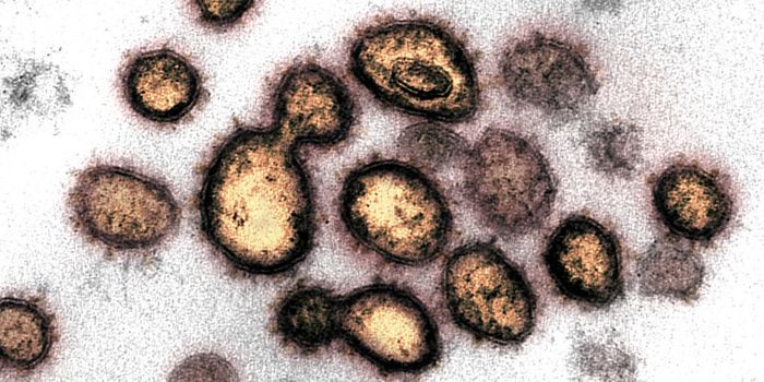
Anthrax is commonly found in soil and can be ingested by animals. It is relatively rare for it to affect people but when it does, it can cost a variety of symptoms and it can be a deadly infection. A three dimensional map of anthrax toxin has now been created by researchers, and the structure may explain how deadly components infect cells and cause disease.
During infection by Bacillus anthracis, anthrax toxin is secreted. There are three parts of soluble proteins that compose the toxin. Similar enzymes make up two parts of it, enzymes called lethal factor (LF) and edema factor (EF); another protein called protective antigen (PA) is the third piece. They all bind together and are taken into cells by vesicles called endosomes. Seven or eight PA proteins subsequently form a channel in the membrane of the endosomes, which lets LF and EF move into the cytoplasm. There, the proteins are able to damage or even kill the cell.
That channel that forms in the endosomal membrane is critical; LF and EF unfold in the lumen of the endosome to be able to pass through the pore. They then are able to reform into their active structures once in the cytoplasm. If you would like to know more about the general pathogenesis of Bacillus anthracic, check out the video below.
The work was performed by a team of investigators led by Robert Liddington of the Sanford Burnham Prebys Medical Discovery Institute in La Jolla and by Isabelle Rouiller of McGill University in Montreal.
It has been published in The Journal of General Physiology.
The team used cryo-electron microscopy to construct a 3D map of the so-called prepore complex that PA and LF form just prior to translocating. The map illustrated seven PA proteins encircling a narrow pore, complete with three LF molecules ready at the rim for translocation. In addition to binding to the PA subunits, the LF molecules also bound to its neighbor. The scientists have suggested that the interactions between LF particles can hold the deadly enzymes in place, preventing them from premature unfolding.
Additionally, as one LF molecule goes into the channel, its neighbor is destabilized such that it can follow behind, going into the pore immediately. That leaves space on the rim of the pore for additional molecules, forming a continuous chain of molecules streaming into the cytoplasm.
Electrophysiological measurements taken by the researchers of the prepore complex support the theory that molecules can be continuously translocated. "We have demonstrated that the anthrax pore can translocate full-length LF in a highly efficient, fast, and robust fashion," explained Rouiller. "The pore can effectively remain fully loaded for extended periods, acting as a conveyer belt while translocating a continuous 'daisy chain' of deadly LF molecules."
For a less technical look at how anthrax affects human health, what it can do to people, and how it is treated, check out the video above.
Sources:
CDC,
Science Daily via
The Rockefeller University Press,
The Journal of General Physiology