Researchers have been looking for ways to image brain activity in animals that are not only alive but behaving normally in a typical environment. There are many hurdles to overcome, and while scientists have been able to create detailed images of brain structure in developing zebrafish, they are not freely moving while the data is obtained. Now, investigators from Helmholtz Zentrum München have created a new way to image neuronal activity in untreated, unrestrained zebrafish, a common model in research. Their invention is also open-source, so it is freely available to anyone. The research has been published in Nature Methods.
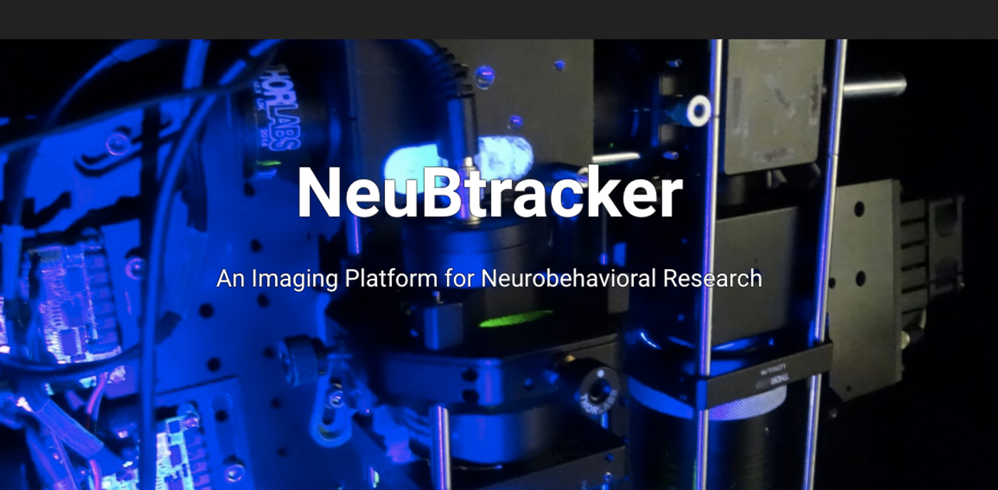
The scientists have created NeuBtracker, which has two cameras; one is for tracking the unrestricted behavior of zebrafish larvae, as the other camera remains trained on the zebrafish head, which is conveniently transparent. Thus, images of the brain can be generated as it's moving.
"This approach makes it possible to observe neuronal activity during unrestrained behavior. We can test the larvae in different environmental conditions and can immediately analyze the effects," explained Dr. Gil Westmeyer of the Institutes of Biological and Medical Imaging (IBMI) and Developmental Genetics (IDG) at the Helmholtz Zentrum München as well as the Department of Nuclear Medicine and Munich School of Bioengineering (MSB) at the Technical University of Munich (TUM).
Zebrafish are great experimental models not only because they are transparent; they also develop rapidly and share sequence similarity with many human genes. Zebrafish can easily be fed different diets, or treated with various drugs, and now the effects of such treatments on the brain can be monitored in a new way.
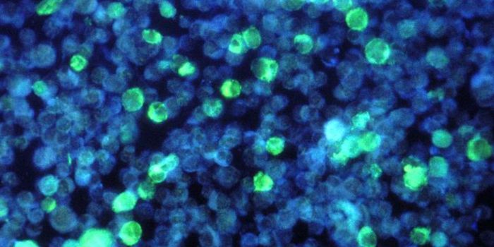
"Now we can finally simultaneously observe the effects of physiologically active substances on the behavior and brain activity," commented the first author of the work, Panagiotis Symvoulidis of the TUM and the Helmholtz Zentrum München. "The selective expression of fluorescent sensor proteins allows us to detect the activity of particular neurons."
"Consequently we can see exactly which areas in the brain are active during specific behaviors," added Dr. Antonella Lauri from Westmeyer's team.
The research team has provided instructions for building the microscope, online, where they are available to everyone. "We wanted to give our scientific colleagues the possibility to build their own NeuBtracker because we had been waiting for such a device for years," Westmeyer explained. "It is finally possible to see the effects of pharmacological substances on the behavior and the neuronal activity -- or other cellular signal processing events -- at the same time and across an entire organism. This systemic approach enables us to make new discoveries and we will, for example, seek to use this device in drug discovery and metabolic research," Westmeyer concluded.
Learn more about zebrafish as a brain research model from the above video, by Howard Hughes Medical Institute.
Sources: Phys.org via Helmholtz Zentrum München, Nature Methods