Anton van Leeuwenhoek (1632-1723) was the first person to make and use a real microscope. He was able to utilize 550 different lenses in order to produce a lens tube that could view objects that were one millionth of a meter, or 270X magnification. Another pioneer of microscopy, Robert Hooke, published Micrographia in 1665 which included illustrations of his observations through the microscope. Despite the progress that has been made since then, technology has lagged considerably regarding the ability to visualize microbial interactions at the macroscale level (mm to cm).
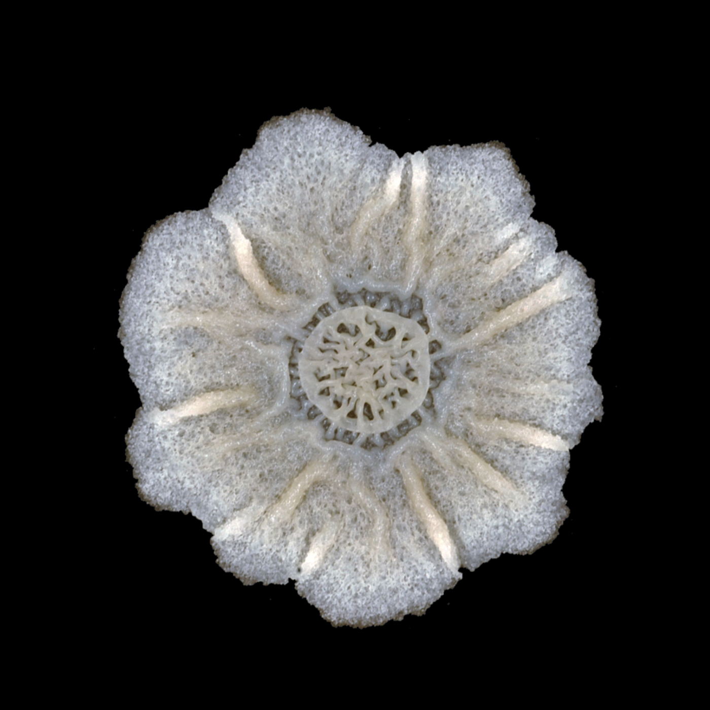
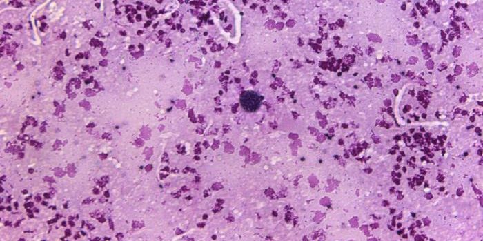
Scientists have developed a MicrObial CHAmber, referred to as MOCHA, which allows for the imaging of microbial communities at the macroscopic level. A colony of bacterial cells is pictured here. Credit: Hera Vlamakis, Harvard University Medical School (Research funded by the Harvard MRSEC, a National Science Foundation MRSEC).
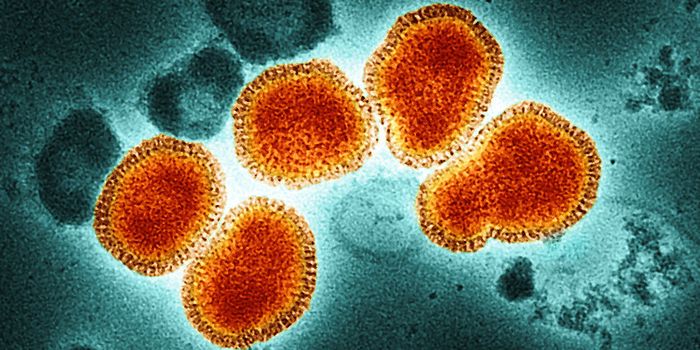
A bacterial colony is defined as a visible mass of microorganisms all originating from a single mother cell and therefore; a colony is actually a group of clones of the bacterial mother cell. The majority of microscopes, such as those developed by Leeuwenhoek, allow for the visualization of a single cell and cannot capture the interactions that occur at the colony level. Because most bacteria exist in communities, it is important to be able to visualize interactions that take place at the colony level, such as those that occur in biofilms.
Until now, the majority of studies of microbial communities at the colony level are conducted using cameras placed within an incubator. This method allows cameras to capture microbial growth over short periods of time however; it has many limitations. This method, which typically involves the use of a camera on a tripod and light source facing down in an incubator, does not prevent dehydration of bacterial growth media (agar). As the moisture in bacterial growth media evaporates, cameras and lenses become fogged up.
In order to overcome this limitation, scientists have developed a specialized microbial chamber that prevents agar dehydration using a double-decker petri dish apparatus. The apparatus is reportedly inexpensive and easy to assemble. It also allows the user to automatically take images of microbial growth on petri dishes at varying magnification, intervals, and duration.
The lower petri dish contains a water reservoir which feeds the upper agar petri dish containing bacteria using a paper wick. A humidifier was placed inside the microbial chamber, removing the lens fog obstacle, and allowed scientists to monitor microbial growth for more than 40 days.
Using this MicrObial CHAmber, named MOCHA, scientists were able to create time-lapse movies of microbial organisms demonstrating the formation of various morphologies on solid agar as well as the liquid-air interface over long periods of time. They were able to create similar movies using fungal microorganisms. Scientists hope that this new technology will facilitate evolution of microscopy for capturing microbial growth at the macroscopic level.
Sources: Journal of Bacteriology, History of the Microscope, National Science Foundation