Have you ever had to say to someone "I'm so bad with names but I remember faces much better."? Well, it turns out the brain has a special region just for recognizing faces. A much-cited study from 1997 by Dr. Nancy Kanwisher, Ph.D. and colleagues from Harvard University describe the neural mechanisms for facial recognition and found that region of the brain: the fusiform gyrus (FG).
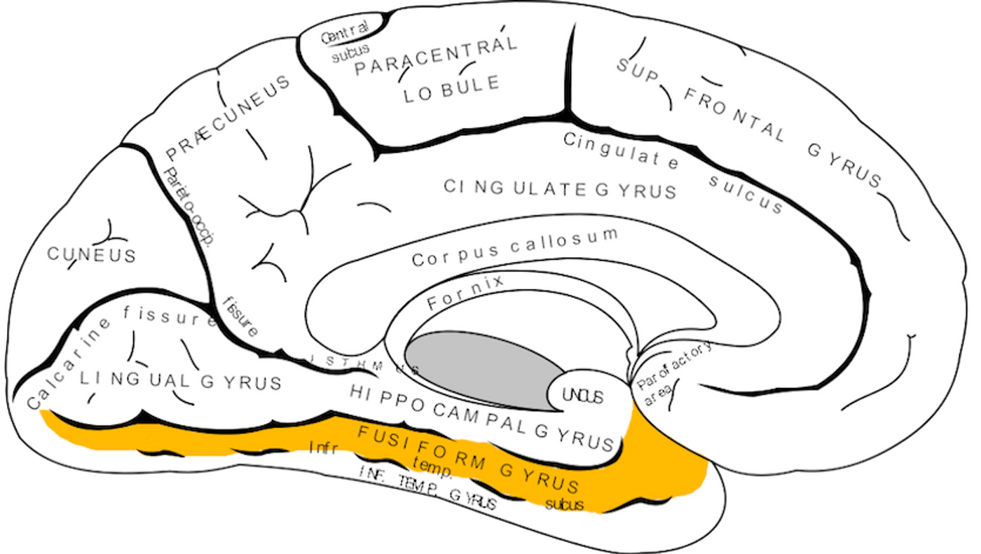
A diagram of the brain seen from the right side of the head (minus cerebellum). Front is on the right.
Photo Source: Wikipedia
Psychological studies had determined that study participants could recognize faces faster than other objects. For cognitive scientists, the faster the recognition, the faster the process. This can be easily demonstrated by the Stroop tests, which shows we read words faster than we identify colors. It appears to be the same with faces. Now enough physiological and imaging evidence has mounted to demonstrate the neuroanatomical characteristics of this phenomenon. Probably the greatest piece of evidence comes from new deep brain stimulation methods that show that when the FG is stimulated faces become distorted.
This is due to the specialized area of the brain that processes faces as a top-down process. That is, the brain receives enough visual information to call something a face, and objects that resemble faces will just be seen as faces. The internet has created many memes for this if you'd like to see for yourself (warning: you will not be able to unsee these "faces").
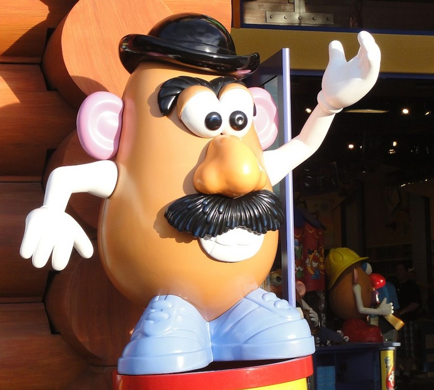
Photo source: PixaBay.com
The relevancy of this brain area today is that dysfunctions in this region could contribute to several neurological diseases: such as autism and mild cognitive impairments (MCI) in the elderly. In terms of autism, in which patients have difficulty with social cues, deficits have been found in this area of the brain. EEG recordings have indicated that children with high-functioning autism may process emotional faces with a decrease in FG processing speed.
Elderly patients with mild cognitive impairment (MCI) may also be suffering from pathologies in this area too. A particular neurological deficit is atrophy of the medial temporal lobe, particularly in rhinal cortices. That is right below the FG. Decreased function of the FG and its surrounding regions may impair facial recognition and, furthermore, is correlated with the development of dementia.
Certain other sensory phenomena have been linked to the FG. Synaesthesia (mixing of perceptions), prosopagnosia ("face blindness"), dyslexia, and facial hallucinations have been proposed to have to do with altered FG processing. In fact, there is a high prevalence of prosopagnosia evident both in children and adults with autistic disorders.
Below is a video explaining the neuroanatomical areas involved in facial recognition:
Video source: YouTube.com
Sources: Journal of Neuroscience, en.wikipedia.org - fusiform gyrus, math.unt.edu, BoredPanda.com, Proceedings of the National Academy of Sciences, CNS Spectrums, Biological Behavioral Research, Science Letter, The Brain and Behavior: An Introduction to Behavioral Neuroanatomy, A Dictionary of Hallucinations, Neuropsychologia, Neuroimage, www.popsci.com