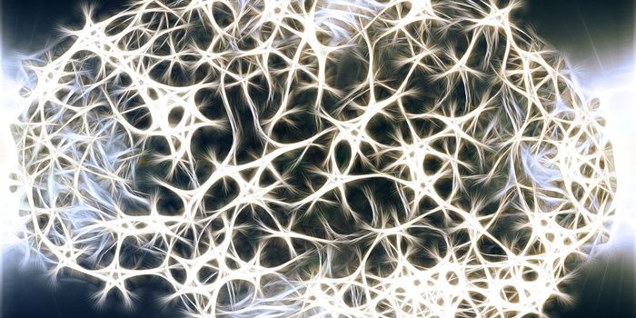
The brain has always been referred to as the super computer of the body. Millions of connections happen just to make a simple move like picking up a spoon or scratching an itch. The cerebral cortex is where most of the action happens. There are approximately 20 billion neurons in the cerebral cortex, and that makes for synaptic connections (intersections of neurons that signal each other) in the trillions. Scientists at the Max Planck Florida Institute for Neuroscience (MPFI) have recently released research about visual processing in the cerebral cortex which shows evidence that the arrangement of synaptic connections within the dendritic field supports an active role for dendrites in how visual information is handled in the brain.
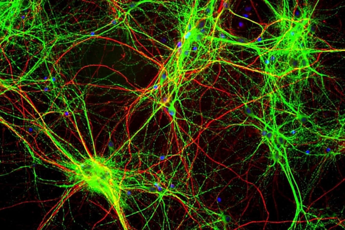
Dendrites are almost like branches that extend out from neurons to complete the synaptic connections that allow neurons to communicate and pass along impulses in the brain. These cortical dendrites are different than others in the body. They have tiny spines that protrude from them and this causes a different kind of synaptic connection. It’s always been believed that these are just the cables upon which the information is passed along in the brain, but the information in the new study from the MPFI suggests that the dendritic network, specifically how it’s configured and arranged actually plays an active role in how visual information is passed along. These networks of dendrites don’t just sit there whilst the information is passed over them, they appear to have a necessary role in coding the information for better processing.
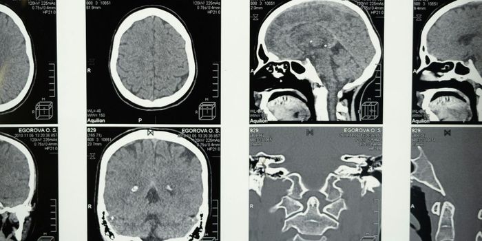
Visual information coming into the brain is complex, so how it’s processed is vital to a person being able to navigate their environment successfully. We know that it’s the neurons responsible for this task, but before the MPFI study, the exact mechanism was unknown because seeing the process was difficult. Brain imaging at this microscopic level is a very new field in neuroscience.
The MPFI team addressed this issue using new microscopic imaging technologies that enabled them to evaluate the input/output functions of individual cortical neurons in the living brain. By using in vivo 2-photon calcium imaging, they were able to comprehend the orientation tuning and spatial arrangement of synaptic inputs to the dendritic spines of individual neurons in the visual cortex of the ferret and compare them to responses in cell bodies and dendritic spines.
Different neurons had different preferences for processing and selecting vertical and horizontal orientation information and the team found that these preferences could be predicted somewhat by adding up the responses within the dendritic spines. But not all the computations could be accounted for in this way. The degree of selectivity in individual neurons was not always exactly related to the whole of the dendritic spine activity. What they found was that spines with similar selectivity were often clustered together along a particular dendrite and the neurons with these clustered arrangements of spines on the dendrites had a greater selectivity. The researchers found that they were able to reliably predict the orientation preference of individual neurons simply by adding up the responses of their dendritic spines. However, the responses of the dendritic spines did not account for the degree of orientation selectivity exhibited by individual neurons. In looking for factors that could account for differences in selectivity, they noticed that spines with similar orientation preference were often spatially clustered along the dendrite and neurons that had a greater number of these clusters exhibited greater selectivity. They also discovered that this functional clustering was correlated with localized dendritic events that are likely to enhance the inputs from the clustered spines. In a sense, spines of a feather, flocked together. Their study was published in the journal Nature Neuroscience on June 13, 2016
The research went further than anything previously has in explaining how the brain processes visual stimuli and orientation from our complex world. The video below explains more about the process, take a look.
Sources:
MPFI,
Neuroscience News,
Nature Neuroscience