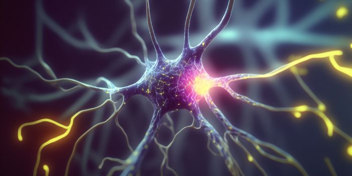
When doctors need to diagnose a brain injury or neurological disease, imaging is crucial to making an informed decision about care. CT scans and MRIs are the norm, but brain imaging and getting a good look at the complex network of neurons and synapses has come up a bit with some high tech features. These sophisticated ways of looking at the brain will ensure that patients get the best care possible after an injury or when treating a neurological condition.
The first is a 7 Tesla or 7T MRI scanner. It produces three-dimensional images of the brain and has a magnet much stronger than most MRI scanners which are between 1.5 and 3 Tesla. Tesla in this case is a unit of measurement, not an eccentric pioneer of electronics. It’s used to quantify the level of flux density (or field intensity) for magnetic fields (also called the magnetic induction). The intensity of a magnetic field can be measured by placing a current-carrying conductor in the field. The magnetic field exerts a force on the conductor, a force that depends on the amount of the current and on the length of the conductor. One tesla is defined as the field intensity generating one newton of force per ampere of current per meter of conductor. Even the lowest level MRIs that are at 1.5 Tesla have very strong capabilities and produce quality images.
The newer 7T machines however get images down to a microscopic level. In human blood there are hydrogen atoms in the water molecules it contains. When they are placed in a strong magnetic field, they align with the field, in a sense acting like a magnet. Radio waves produced by the scanner excite these hydrogen atoms causing them to spin Once they stop twirling, they emit specific signals that are detected by the scanner and processed by computers to produce an image. With the 7T scanners, even the tiniest fluctuations in blood flow in the brain and other metabolic changes are picked up and this makes the images much more helpful to doctors.
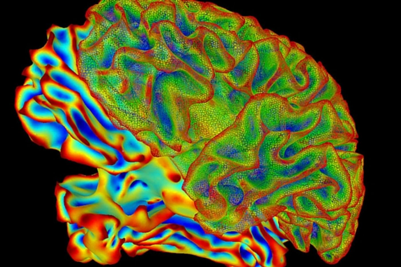
Another advance in brain imaging comes from taking MRI images and images from Diffuse Tensor Imaging scans, which track the movement of water molecules through the brain’s fibrous stuctures, and creating a 3D holographic image of the network of fibers in the brain. Being able to visualize these structures is important for evaluating patients who may have Alzheimer’s or other forms of dementia. The British company Holoxica has created a prototype of the technology and hopes to have it in production soon. The CEO Dr. Javid Khan said in a press release, “A recent study concluded that 3D visualisation is up to 75 per cent better than 2D for certain types of tasks including spatial manipulation, finding, identifying or classifying objects, but our future plan is to take 3D imaging to the next level using our holographic video display, which is being designed for medical imaging machines including MRI, CT and Ultrasound scanners.” The video below explains these two kinds of imaging and what they mean for neuroscience.
Sources:
Holoxica,
The Daily Record,
Siemens