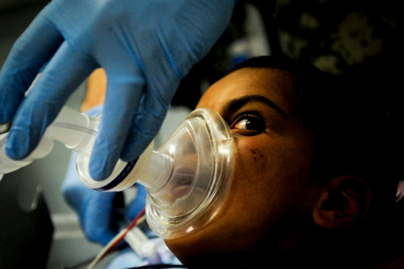
General anesthesia carries many risks with it, as does any surgical procedure. The effect that anesthesia medication has on the body and particularly the brain isn’t completely known. Different patients will respond in different ways to medications and close monitoring during surgery is crucial. The drug Propofol made headlines after being used by Michael Jackson for insomnia, as opposed to surgery. Autopsy reports on Jackson showed that he died of an overdose of the drug. While it’s still used safely during supervised surgeries, finding out how the drug acts on the brain will increase awareness of how best to use it in a hospital setting and hopefully decrease its use outside of the OR.
Anyone who has had surgery can attest that you are seemingly awake one minute and unconscious the next. In the cortex of the brain however, it doesn’t work exactly like that A new study by researchers at Massachusetts General Hospital and Harvard Medical School shows that brain activity gradually shifts rather than shutting down immediately.
Most researchers have looked at the effects of anesthesia using EEG readings. These recordings of the electrical activity in the brain do show some effects of anesthesia, but it’s not a perfect method. The study done in Boston used Macaque monkeys who had sensors implanted directly in the brain tissue and these sensors were able to show much more detail, literally down to specific neurons, than an EEG would have revealed. The monkeys were trained to perform a task that would indicate alertness and were tested for responses to tactile and auditory stimuli. The researchers looked at brain activity while they were fully awake and as they lost consciousness to show how the brain was functioning.
Looking at two areas in the brain, one that deals with incoming sensory information and one involved with movement, the team saw that these areas were working together in sync before the administration of Propofol. At the point where the monkeys were just beginning to lose consciousness, activity in the two areas were no longer working in tandem, but rather each area showed its own patterns. At the point of being fully asleep, there was a surge in the nerve cell activity of the area of the brain responsible for movement and a similar surge a few minutes later in the area responsible for processing sensory input. Finally when the monkeys were full anesthetized by the drug, the two areas of the brain synced up again, working much like they had before the drug was administered.
In an interview with Science News, the co-author of the study, Yumiko Ishizawa of Harvard Medical School and Massachusetts General Hospital expressed surprise that period between being fully awake and fully asleep had shown such different nerve cell activity. More than anything, the study showed that the effects of this drug, and likely other drugs used in anesthesia, is quite a bit more complicated. The brain, and thus the patient, doesn’t simply go from awake to asleep as if there was a switch that was flipped, but rather the process is gradual and results in specific patterns in different parts of the brain. The video below talks more about the process of anesthesia and this study of it.
Sources: Science News, Journal of Neuroscience