The spinal cord is the pathway between the brain and the rest of the body. Much like a super highway, messages for muscle movement, bodily functions and so much more have to pass along this this vital part of the anatomy. That is why it’s so devastating when a spinal cord injury occurs. Neurologically, depending on the injury and it’s location, much of a person’s body can be affected. Paralysis, a loss of sensation and function go along with severe spinal cord injuries.
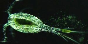
As yet, medical science has not figured out how to regenerate the spinal cord and restore function, but a team at Duke University might have advanced the cause just a bit further with their recent study. How? They have a tiny little aquatic friend to thank for the new information, the freshwater zebrafish. Zebrafish are used extensively in research since they have about 25,790 protein-coding genes.
Roughly 70% of these are genetically related to what humans have, so they make for good research animals. They are also vertebrates and can repair their own spinal cord. Researchers at Duke wanted to know how this happened and the study they recently published could lead to new treatments to repair tissue.
Senior investigator Kenneth Poss, professor of cell biology and director of the Regeneration Next Initiative at Duke said in a press release, “This is one of nature’s most remarkable feats of regeneration. Given the limited number of successful therapies available today for repairing lost tissues, we need to look to animals like zebrafish for new clues about how to stimulate regeneration.”
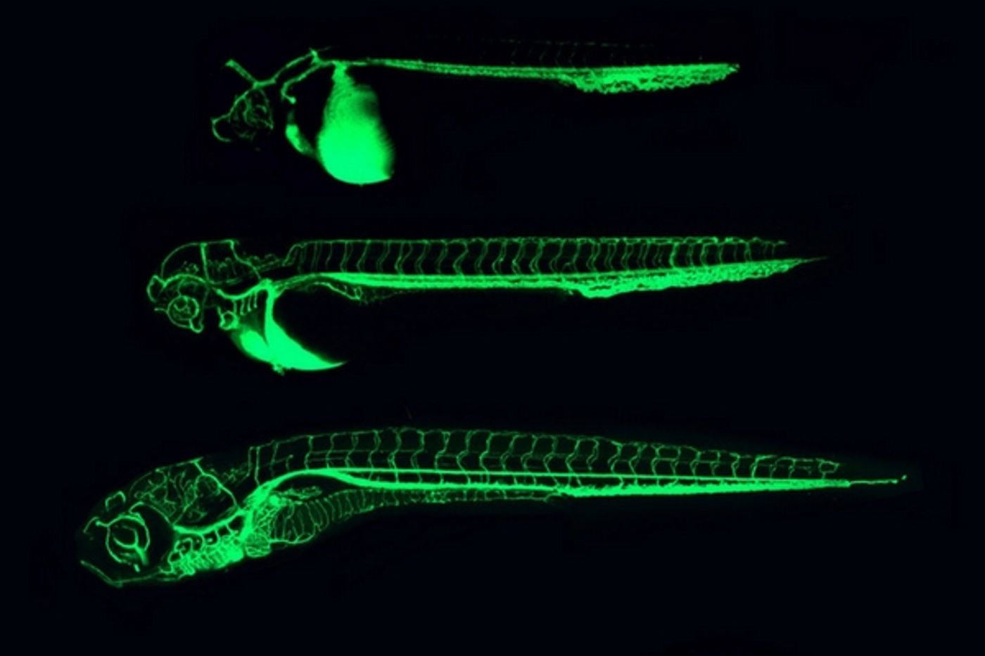
The spinal cord in the fish regenerates by forming a bridge of cells. While it takes about 8 weeks for the fish to repair a severed spinal cord, when it’s complete, paralysis is completely gone and function is restored. When regeneration first begins, cells extend projections of matter outward, sometimes at distances tens of times their own size. The injury site is like a wide chasm that has to be knitted together. Once the nerve cells join in the process, the paralysis is reversed. This kind of regeneration happens at the molecular level and that is what the Duke team was able to identify.
They looked at several genes that were activated by an injury and of those, seven coded for certain proteins that were secreted from cells. The most significant one, CTGF, or connective tissue growth factor, increased dramatically after the injury. It was this protein that drove the first cells at the injury site to start bridging the gap. Fish that had been modified genetically to eliminate CTGF could not regenerate their spinal cords at all.
Humans have this protein as well, however the team did not think that in mammals, CTGF would be enough to trigger regeneration, mostly because of how humans form scar tissue near injuries. They hope to expand the research into mouse models, which would show how the CTGF behaves in an anatomy more similar to humans. The human form of CTGF is only about 90% similar to that of the fish, but the team is still hopeful that looking at how this protein is expressed in other species will lead to more information on spinal cord regeneration and other nerve injury. The video below, from Duke, explains how this research could be a new beginning for neurological disease and injury.
Sources:
Duke University,
Science Magazine,
Pain News Network