Brain development, what we are born with, what develops during childhood and adolescence, and what is lost as we age are big concepts in neuroscience research and many studies look at each of these phases. It’s known that humans are born with more neural connections and synapses than are actually needed. As childhood progresses into the teen years, many of these connections, like overgrown trees or bushes, are pruned in the brain’s maturation process. It’s been thought that once this pruning is complete, the brain is complete and remains at a stable level. That isn’t actually the whole picture; it’s a bit more complex. Two papers recently published by the same senior author, show that certain brain regions have a very specific cellular composition and that there is microscopic tissue growth that results in structural and functional changes in the part of the brain that recognizes faces.
The two studies, conducted at Stanford and published in the January 6, 2017 issue of Science and the Nov 30, 2016 issue of Cerebral Cortex, looked at the brains of children, after the period of initial pruning and found that the brain does not stay the same until aging sets in. There is more growth and this growth, of very specific cells, mirrors the improvement of facial recognition that happens as children grow up.
Kalanit Grill-Spector, a professor of psychology at Stanford and senior author of both papers explained, “I would say it’s only in the last 10 years that psychologists started looking at children’s brains. The issue is, kids are not miniature adults and their brains show that. Our lab studies children because there’s still a lot of very basic knowledge to be learned about the developing brain in that age range.”
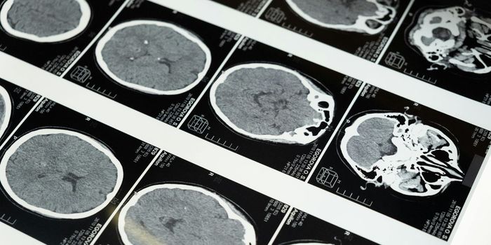
In examining the brains of children the two concepts that became clear were the cellular make up of the region of the brain that processes facial recognition and the growth of tissue in that region that continues after the initial pruning of unnecessary neural connections. In MRI scans of the children, the team was actually able to see new tissue growth. Jesse Gomez, a grad student in the lab and lead author of the paper in Science stated, “We actually saw that tissue is proliferating. Many people assume a pessimistic view of brain tissue: that tissue is lost slowly as you get older. We saw the opposite – that whatever is left after pruning in infancy can grow.”
As for the cellular composition of this part of the brain, that was more difficult to actually see. The team at Stanford collaborated with the Institute of Neuroscience and Medicine, Research Centre Jülich, in Germany, who obtained thin tissue slices of post-mortem brains. For a year the two groups matched the functional MRI scan results to corresponding brain slices and were able to extract the actual microscopic samples of the cell structure. Naturally this cannot be done with living subjects, so the combination of post-mortem tissue and living MRI data is what made it possible to understand the cellular structure of this part of the brain.
The scans used were not simple MRIs either. The team used a combination of functional MRI (fMRI) scans that measured brain activity while the test subject is completing a mental task and quantitative MRI scans which measure the proportion of tissue to water in the brain. This scan has been used to show changes in the fatty insulation surrounding the long neuronal wires connecting brain regions over a person’s lifetime, but the team at Stanford was the first to use it to measure cell growth. It was this growth that was responsible for tissue differences from childhood to adulthood in the part of the brain that recognizes faces.
Grill-Spector summed up the work by saying, “If you had told me five or 10 years ago that we’d be able to actually measure tissue growth in vivo, I wouldn’t have believed it. It shows there are actual changes to the tissue that are happening throughout your development. I think this is fantastic.” Check out the video to see more about this new information.
Sources: Stanford University, Cerebral Cortex, Science