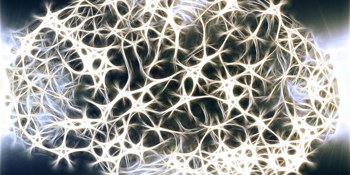
Neuromodulation. Isn’t that a pretty cool word? Sounds like something Marvin Martian would say, but it’s no cartoon, it’s a very real part of neuronal communication in the brain. When the neurons in the brain communicate, it involves a fairly orderly process of molecular neurotransmitters traveling over intersections of nerves called synapses on the way to stimulate other neurons. This is how different parts of the brain interact to accomplish different tasks. It’s not always that efficient however, sometimes parts of the brain get flooded with neurotransmitters and neuromodulation, the way the brain regulates this flow, and in turn determines mood, is triggered.

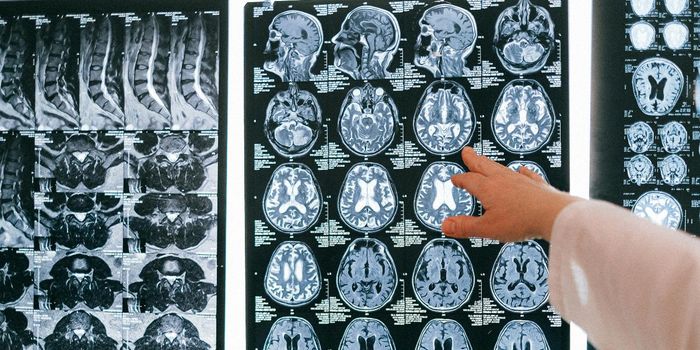
In terms of certain brain disorders or mental illnesses, understanding neuromodulation is a crucial part of finding ways to treat disease. Visualizing it has been one of the most difficult parts of this task, but scientists at Hyungbae Kwon’s lab at Max Planck Florida Institute for Neuroscience (MPFI), and collaborators from University of Geneva, Korea University and Max Planck Institute of Neurobiology have developed a process that involves gene expression and allows neuromodulation to be observed in real time.
The team at MPFI has built on some previous work and come up with the Inducible Tango technique, called iTango. Initially, the technique from a decade ago would cause neurmodulators to express a green fluorescent protein, so researchers could identify them. This was of limited benefit however, because the fluorescence would just stay on and spread to more neurons, regardless of whether or not they were being stimulated by neurmodulators.
iTango is much more specific because it involves researchers shining a blue light on whichever neuron they want to observe. In the new method, the glow of the protein only happens if a certain neuromodulator is present. When the light is turned off, the protein ceases to fluoresce. It’s a significant advance in molecular imaging because researchers now have what amounts to a “light switch” that gives them control over the timing of their observations and the specific neurons they want to investigate.
RELATED: Sound Waves for Essential Tremor
In this study where the process was developed, the team used iTango to identify two groups of neurons in mice. One that regulated movement and one that was involved in reward circuitry. Not only were they able to observe these processes in the mouse model, they went even further and were able to induce and inhibit certain behaviors using the light. The mission of Dr. Kwon’s lab is mostly neuroscience research, the formation of brain circuits and how they influence disease, mood and brain function, but the iTango process could have applications in drug development and targeted cancer therapies as well. Still, the team at MPFI is excited about the neuroscience applications and how the technique will aid in brain research. In a press release, Dr. Kwon explained, “The most important part of this story is that we made this technique light sensitive. The light sensitivity provides a huge advantage because you can apply it to basically any signaling. iTango will be useful for visualizing and manipulating the neuronal circuitry that underlies drug-induced behaviors and psychiatric diseases related to neuromodulation, such as mood disorders or schizophrenia.” Take a look at the video below for an explanation of how it works.
Sources: Max Planck Florida Institute, Nature Methods, Oxford Index