Look around at where you are right now. How will you get home? How will you get to work or the coffee shop or the gym? You will get around because your brain has specialized neural networks that process your environment and encode it in your memory. It was this sort of research that earned three neuroscientists a share in the Nobel Prize for Medicine in 2014. These networks are essentially the brain's GPS system. When they are disrupted, as is often the case in patients with cognitive decline (from dementia, Alzheimer's disease, etc.) we can lose our way.

There is new research, however, from the Flanders Institute for Biotechnology, that shows neural activity in a region of the brain not previously believed to be involved in spatial memory and navigation. The area is the retrosplenial cortex and knowing that it's involved in memory and environment could be significant in finding therapies for patients who suffer from memory deficits.
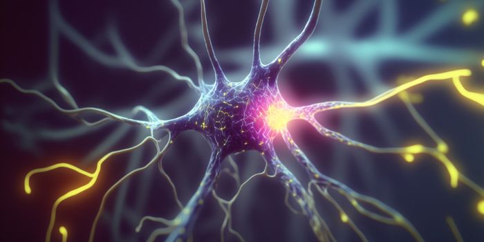
Most of the information in spatial memory is found in the firing of neurons in the hippocampus. The hippocampus is memory central for the brain. In animal models, cells in this area, called place cells, fire when entering an environment that is known. It's an efficient process, just a small group of cells, which allows for the rest of the brain to store new memories. But as the most recent research shows, it's not the only area of the brain that is active in learning and orientation.
The retrosplenial cortex also has networks of neurons that fire, and is a connector between the hippocampus and other areas of the brain like the visual cortex. In some studies, patients with AD have damage to this part of the brain, which accounts for why some people with this form of dementia often get lost or forget how to perform daily tasks.
Professors Vincent Bonin and Bruce McNaughton at Flanders along with colleagues Dr. Dun Mao and Steffen Kandler wanted to understand the role of this part of the brain, so they studied mice who were moving on a treadmill that was equipped with tactile stimuli. They used sophisticated genetic techniques to label the neurons that fired as well as microscopic real-time imaging to compare the network activity in the retrosplenial cortex to that in the hippocampus.
Prof. Bonin explained, "Previous studies could only record from a few retrosplenial neurons simultaneously. With our cellular imaging technique, we could monitor the activity of hundreds to thousands of neurons simultaneously, which gave us a rich view into the neurons' activity patterns."
What they saw was an entirely new set of neurons in this cortical area, with patterns that had not been seen before and which responded differently to stimuli than the place cells in the hippocampus. Now that this area's involvement in spatial memory and its unique firing patterns have been uncovered, the team hopes to conduct further research into how exactly the two areas (the hippocampus and the retrosplenial cortex) interact. Knowing precisely how these two regions are connected is likely a key factor in how neurological disorders that cause dementia occur and how they might be treated. The video below has more information, check it out.
Sources: Flanders Institute for Biotechnology, UPI, Nature Communications, Nobel Prize.org