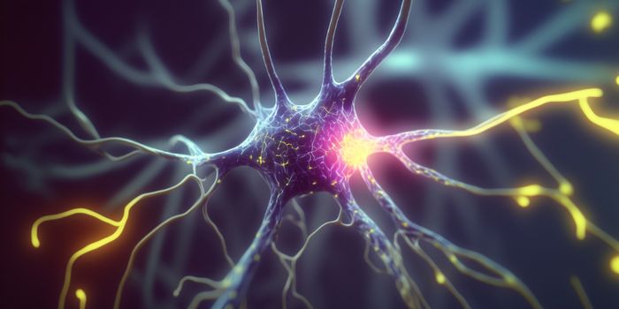
As any neuroscientist will undoubtedly attest, if there were a way for researchers to be inside a brain, able to visualize the cells, see how they work and interact with them, that would be a most fantastic adventure.
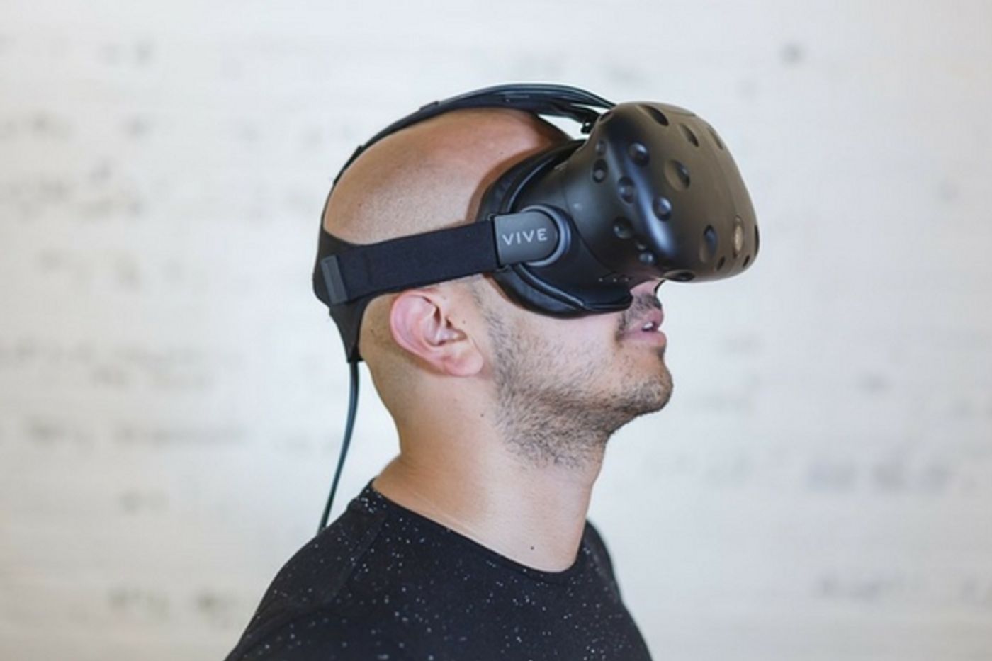
As it happens, something much like that is now possible. Researchers from the Wyss Center for Bio and Neuroengineering collaborated with experts at the University of Geneva to develop a truly immersive virtual reality environment. By donning a pair of VR glasses, users can, at least virtually, be inside a brain. Cellular behavior can be seen and whoever is wearing the goggles can interact with the brain environment. It's a mouse model, but the detail is incredibly true to life, and mouse models are often used in neuroscience research.
A poster about the project was presented at the Society for Neuroscience 2017 meeting in Washington DC recently. So how was the brain environment created? With vast amounts of data. Because of advances in neuroimaging, researchers can now see individual neurons and their firing patterns. The Wyss Center has a light-sheet microscope, and it's one of only three such pieces of equipment in the world.
It was built by Dr. Stephane Pages, a staff scientist at the Wyss Center and a principal researcher at Campus Biotech at the University of Geneva. Called the Clarity-Optimized Light Sheet Microscope (COLM) it's capable of looking at a tiny slice of brain tissue and creating multiple images, like slices, and from that, a 3D rendering can be made. The COLM can eliminate the opaque fluid found in tissue samples, and this allows an entirely transparent image to be formed, in 3D.
The VR component is helpful because the data and imaging that come out of such advanced equipment can be hard to manage. While the structures visualized are tiny, the amount data needed to fully utilize the images is enormous. Dr. Page explained, "The immense data volumes produced by today's high-performance microscopes are driving the development of new methods to visualize the brain. We have developed this virtual reality system to reconstruct cellular level neuroanatomical data in 3D space. The system provides a practical solution to experience, analyze and quickly understand these exquisite, high-resolution images."
The images detailed at SfN2017 were even able to show the neuronal pathways right down to the dendritic spines which reach out of a neuron to pass along electrical and chemical signals throughout the brain. The team at Wyss has been able to combine the imaging of the COLM with data analysis tools to boil all the images and metadata down to something that is recognizable. While wearing the VR headsets, users have access to handheld digital pointers that can interact with the data in real time, allowing them to zoom in on structures, slice through some images and see the brain at work. In the future the team hopes the technology can be used in training surgeons, and developing neurotechnology. Take a look at the video below to see what it's like being inside the brain.
Sources: Wyss Center, University of Geneva, Terra Daily