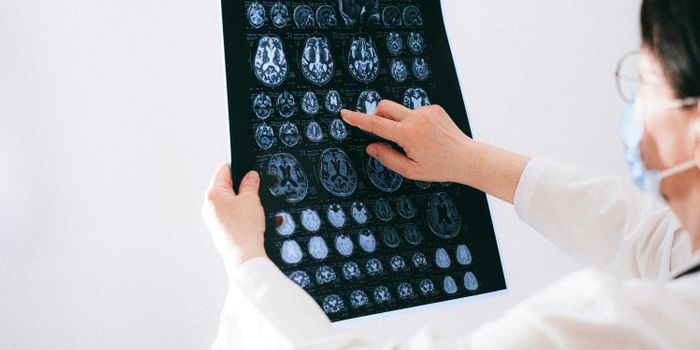
Mental illness is sometimes looked at as a character weakness or something a person just has to "shake off." New research using magnetic resonance imaging (MRI) of the brain shows that there is a specific pattern of structural anomalies in the brain tissue of patients who have a diagnosis of depression and social anxiety. These findings could be a significant breakthrough in not only the treatment of mental illness but also the end of the stigma that some face when trying to recover and treat depression and anxiety.
The two conditions that were examined in the research from scientists at Sichuan University in Chengdu, China were major depressive disorder (MDD) and social anxiety disorder (SAD) The CDC estimates there about 16 million people in the United States who suffer from MDD, and many more who less severe forms of depression. Patients with MDD struggle daily just to be active and involved in their jobs, families, and communities.
Social anxiety disorder (SAD) can also be debilitating and prevent someone from enjoying friendships, a social life or even daily activities that involve being out in the community among people. MDD and SAD share some of the same symptoms, and it's not uncommon for a patient to be diagnosed with both. Many research studies have looked at the theory of whether or not the two disorders might have the same underpinnings in the brain. In the most recent research, the two conditions were examined regarding structural components.
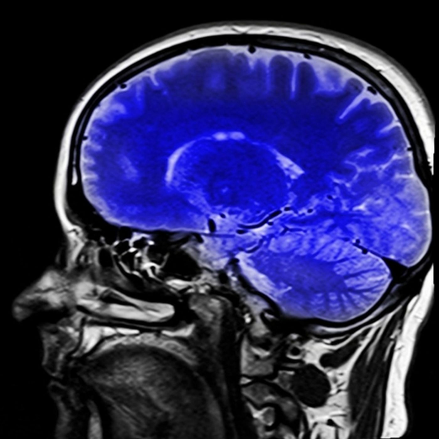
The team at Sichuan University examined 37 patients with MDD, 24 with SAD and 41 healthy participants as a control group. The imaging looked specifically at how thick the cortex, which is the outer layer of the brain, was in each group of patients.
In the groups of patients with MDD and SAD, there were grey matter differences that were not shown in the healthy study participants. The abnormalities were most prevalent is the salience and dorsal attention networks. Both of these brain regions are involved focus, attentiveness and processing stimuli in order of importance.
To function appropriately in social situations, work, and relationships, the brain needs to sort out all the stimuli in our environment. This process is often disrupted in patients with MDD or SAD. In the study results, the patients who had a mental health diagnosis had thicker grey matter in the insular cortex, compared to the healthy controls. The insular cortex is an area that is heavily involved in perception, social skills, and self-awareness.
Youjin Zhao M.D., Ph.D., from Sichuan University was the lead author of the study and explained, "Our findings provide preliminary evidence of common and specific gray matter changes in MDD and SAD patients. Future studies with larger sample sizes combined with machine learning analysis may further aid the diagnostic and prognostic value of structural MRI."
The team hopes to continue the research with a more substantial trial group and make use of machine learning to assess the data collected. The study was presented at the 103rd Scientific Assembly and Annual Meeting of the Radiological Society of North America in Chicago last month. The video below has more information on what researchers were able to determine from the MRI scans.
Sources: Radiological Society of North America, Diagnostic Imaging, iNews