
Assistant Professor, Department of Obstetrics Gynecology and Reproductive Biology, Institute for Quantitative Health Science and Engineering, Michigan State University
BIOGRAPHY
Insights from Imaging the Implanting Embryo and the Uterine Environment in Three Dimensions
Presented at:
Cell & Developmental Biology Virtual Meeting
Speaker
Abstract
Although much is known about the molecular maternal-fetal interactions during implantation, the 3D architecture of the uterine environment in which the early embryo develops is not well understood. Using confocal imaging in combination with 3D analysis we identified dynamic changes in the luminal and glandular structure of the murine uterus in preparation for implantation. Further, we used this methodology to uncover patterns and mechanisms of pre-implantation embryo movement in the uterus. Our analysis reveals three distinct pre-implantation stages: a) Embryo entry; b) Unidirectional movement of embryo clusters; and c) Bidirectional scattering and spacing of embryos. We show that the unidirectional movement of embryo clusters is facilitated by a mechanical stimulus of the embryo as a physical object and is regulated by adrenergic uterine smooth muscle contractions. Surprisingly, we find that embryo scattering, is independent of muscle contractions but instead relies on LPAR3 signaling mediated embryo-uterine communication. Our data supports a model where murine uterine implantation sites are neither random nor predetermined and are a function of the number of embryos entering the uterine lumen. We propose that the presence of embryo clusters in the uterine horn provides an opportunity for the uterus to sense and count the embryos, followed by scattering and even spacing of these embryos along the given length of the horn. These studies have implications for understanding how embryo-uterine communication is key to determining an optimal implantation site, which is necessary for the success of a pregnancy.
You May Also Like
FEB 17, 2026 | 8:00 AM

In cell therapy applications, commonly employed cell purification platforms for large-scale isolation of immune or stem cells separate cells based on a single cell surface antigen. Enrichmen...
FEB 18, 2026 | 8:00 AM

With advancing food allergy research and emerging therapies, new allergy testing requirements arise. In this context, Basophil Activation Testing (BAT) has emerged as an essential component...
FEB 18, 2026 | 1:00 PM

Energy-efficient filtration solutions that reduce hidden chemical exposure from benchtop work and lab equipment to improve overall laboratory safety...
FEB 19, 2026 | 7:00 AM
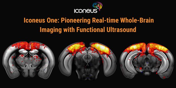
Discover how Iconeus One, the premier functional ultrasound (fUS) system for neuroscientists, is transforming brain function studies. Based on ultrafast plane-wave sonography, Iconeus One of...
FEB 19, 2026 | 8:00 AM

The collection of high-quality genomic DNA remains a major barrier in pediatric and neurodevelopmental research, particularly among children with autism spectrum disorder (ASD) and other neu...
FEB 23, 2026 | 9:00 AM
C.E. CREDITS

Explore decentralized liquid biopsy with SOPHiA GENETICS and MSK-ACCESS®, expanding access, improving outcomes, and accelerating precision oncology....
Loading Comments...