Precision medicine is an emerging field where clinicians tailor treatment plans for each individual based on their genetic history, lifestyle, and environment. Taking this approach one step further, researchers have developed mini-model of the human body and cancer that can predict the route of metastasis and which drugs may prevent this from occurring.
The system, known as “
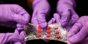
metastasis-on-a-chip”, is the first laboratory invention to model cancer spread in 3D. It was developed by researchers at the Wake Forest Baptist Medical Center's Institute for Regenerative Medicine, who published their findings in the latest issue of Biotechnology and Bioengineering.
"We believe the metastasis-on-a-chip system has potential for making meaningful advances in cancer investigation and drug discovery," said Aleksander Skardal, lead author of the study. As the first proof-of-principle study, the team focused on metastasis from the colon to the liver, a common route seen in invasive colorectal cancer.
To model cancer migration in 3D, researchers first needed to mimic organs and blood flow, as in a living system. They took human cells from the intestines and liver and encapsulated these in a gel-like substance to create two distinct mini organoids held in independent chambers. The device holding the organoids had etched micro-channels through which researchers simulated blood flow. These channels also provided a means for testing drug treatments.
By tagging the cancerous colorectal cells with a fluorescent protein, the researchers could track the cells’ migration in real time. With highly invasive colorectal cancer cells, researchers observed tumor growth in the colon organoid and subsequent metastasis to the liver organoid via the circulatory system. Metastasis was not observed in a non-invasive colorectal cell line.
Metastasis can be inhibited by some anti-cancer drugs. To test this intervention in the chip system, the researchers introduced Marimastat and 5-fluorouracil in two separate trials. They found Marimastat led to confinement of the tumor in the primary colon organoid, while 5-fluorouracil reduced the cancer cells’ metabolic activity.
So far, the team is impressed with how the device mimics responses in patients. They plan to test more compounds to assess whether the same human effects can be gleaned from the chip system. "If this link can be validated and expanded, we believe the system can be used to screen drug candidates for patients as a tool in personalized medicine. If we can create the same model systems, only with tumor cells from an actual patient, then we believe we can use this platform to determine the best therapy for any individual patient," said Skardal.
While this 3D cancer modeling is a huge advancement over 2D systems in petri dishes, much more work has to be done before “metastasis-on-a-chip” can realistically mimic a human system. For example, many organs and multiple pathways are often involved in real metastatic events. Thus the model has to be able to incorporate these real complexities to provide meaningful calculations about cancer response and migration. The team plans to address this by adding more organoids, such as the lung and bone marrow. Additionally, they also plan to add a layer of endothelial cell barrier in channels of the device, to mimic a more realistic circulatory system.
Researchers hope their reductionist approach to studying metastasis has the potential to advance cancer investigation and drug discovery, providing more tailored and precise treatments for cancer patients.
Additional source:
Wake Forest press release