Scientists at the Dana-Farber Cancer Institute in Boston, Massachusetts are launching a cancer tissue repository for use in creating patient-derived tumor mouse models. The initiative, called the
Public Repository of Xenografts (PRoXe), aims to reduce the high failure rate of drugs in early clinical trials.
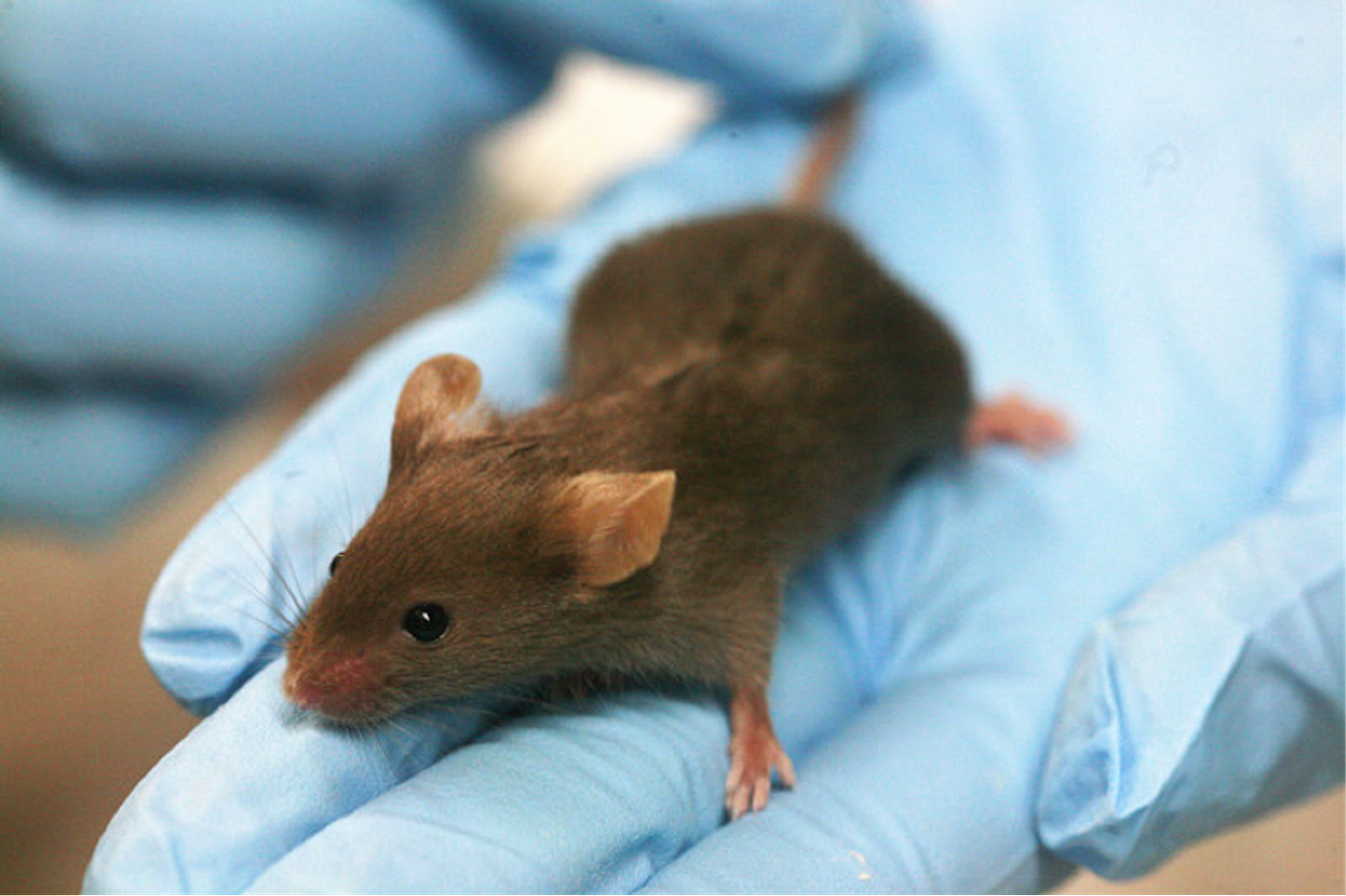
The technique of using mice to model human-derived tumors is not new. Officially known as Patient-Derived Xenografts (PDX) — from the Greek “xenos” meaning “foreign”— the technique has been revolutionizing cancer research since it was first described in 1988. More recently, PDX mice have been referred to as personalized mouse “avatars.” These animals have actual tumor samples from patients implanted in them, and as the tumor cells grow in the mouse avatars, the researchers can test for new mutations, identify different targets, and test different combinations of drugs to halt tumor progression.
"About 90 percent of compounds that show anti-cancer activity in pre-clinical tests don't work when given to patients. By trying drugs in PDX models, we can 'mimic' large and expensive human clinical trials and get answers about efficacy more quickly, less expensively and without the need for patients to get investigational drugs that won't work," said David Weinstock, a physician-scientist clinician at Dana-Farber, and senior study author.
The aim for the repository is to have a centralize location to house many different types of cancer tissues. This open-source repository is available to researchers worldwide, who can request frozen samples to be transplanted to mice in their own labs in order to test different drugs. The PRoXe already contains biopsy material from bone marrow, peripheral blood, and lymph nodes of the mice.
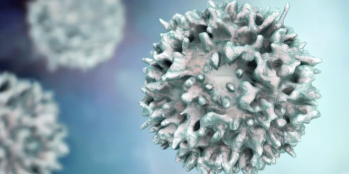
"Using this repository, we demonstrate that large studies of acute leukemia PDXs that mimic human randomized clinical trials can characterize drug efficacy and generate transcriptional, functional, and proteomic biomarkers in both treatment-naive and relapsed or refractory disease," the authors wrote.
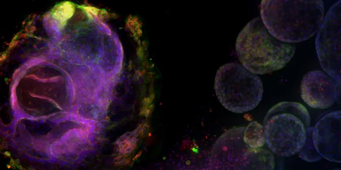
Mouse models are essential to medicine and research, as they allow researchers to explore many biological processes that are otherwise impossible to do in people. Moreover, studying tumor biology in the context of a living mammal provides much more accurate information than using cell lines, which completely lack the systems interactions component. And while researchers are developing newer ways to study and treat cancer in 3D artificial systems, mouse models still remain the gold standard for providing answers that inform doctors on the best course of action for human health and disease.
"These studies allow us to analyze what a drug is doing in many phases of treatment," said Elizabeth Townsend, the study’s co-first author. As a proof-of-concept study, the team demonstrated the applications of PDX mice for an experimental drug in PDX models of leukemia with and without a mutation in the TP53 gene. They showed mice without the mutation responded better to the drug, offering evidence for testing the drug in leukemia patients with similar genetic status.
Elsewhere, other researchers are also using PDX mice in their studies. One used mouse models as
personalized “avatars” for human melanoma cancer to find the right targets and drug combinations to successfully stop tumor growth. The team at Dana-Farber hopes to collect these models and expand the power of the PDX mouse in treating cancer.
Additional source:
Dana Farber press release