How do heart cells follow their genetic programming to create a heart? What can mice hearts do to help scientists understand more about the innerworkings of the human heart? Harvard Medical School scientists explore these questions and more, with their new study of embryonic cardiac development in mice.
“The hope is that now we’re beginning to understand at a single-cell level how perturbations in genes and cells lead to changes in cardiac structure and clarify what the important steps are for how the heart is built,” said cardiology researcher Jonathan Seidman, PhD. Seidman and the team from Harvard began their study of genes and heart cells with samples from seven completely different stages of embryonic development in mice.
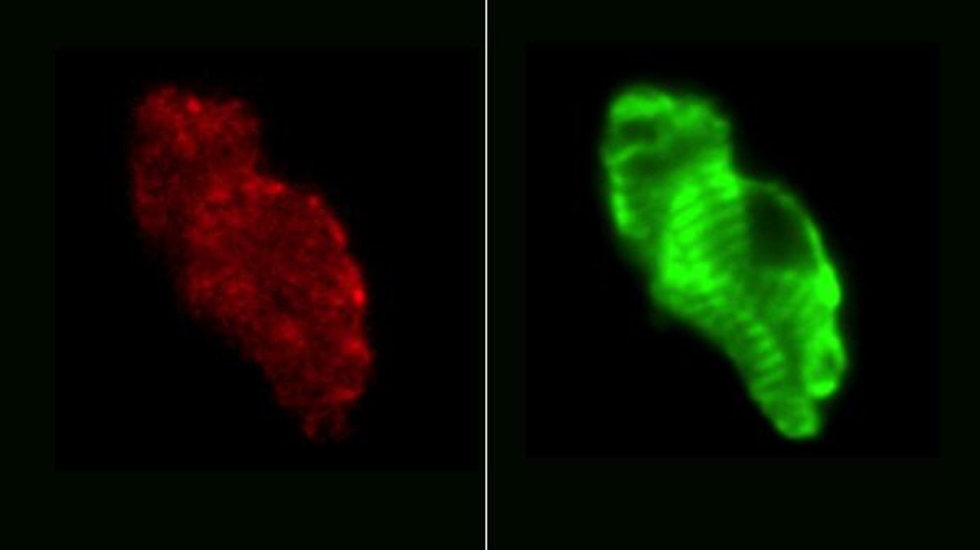
Using single-cell RNA sequencing techniques, researchers divided up the cells from each sample into three known categories:
- Myocardial cells (heart muscle cells)
- Endothelial cells (make blood vessels)
- Fibroblasts (“hold everything together”)
For each cell type, they watched the changes in genetic activity over time; this study was the first time scientists have looked at a “map of the heart” with 100-cell resolution.
“The study provides both a temporal and spatial atlas that plots the development of the different cell populations in each of the four chambers,” Seidman explained. “The converse is also true: by looking at a cell’s RNA expression, we can gather clues to its origins.”
The results showed errors in specific cell types in mice with a genetic mutation also associated with congenital heart defects in humans, as well as a plethora of information concerning the location of certain cell types in the heart at different times during development. However, they also made an unexpected discovery: a seemingly new cell type that express genes from both myocardial cells and fibroblasts. Researchers are still unsure what these cells are and what role they play in embryonic development.
Mouse hearts are similar to human hearts and make a good model for study, but several differences limit the information scientists can glean from mouse studies. Mouse hearts are three thousand times smaller and beat ten times as fast as human hearts. But shared similarities between the two structures like four heart chambers, electrical signaling, and molecules involved in muscle function make the mouse heart just good enough to take away significant conclusions.
Seidman and the team from Harvard plan on continuing research with mice heart structure and function, looking for concrete explanations for why certain congenital heart defects develop. The present study was recently published in the journal
Development Cell.
Source:
Harvard Medical School