One explanation for why heart failure develops goes all the way to the miniscule organelles inside individual cells. From Georgia State University, researchers from a new study show how one mitochondrial protein, FUNDC1, is connected to heart dysfunction and, ultimately, heart failure.
Understanding FUNDC1
FUNDC1 is an outer mitochondrial membrane protein whose function has yet to be entirely explained. What scientists from the current study do know, though, is that reducing its levels in heart muscle cells, called cardiomyocytes, initiates cardiac dysfunction and makes existing dysfunction worse.
Additionally, intervention between FUNDC1’s interaction with certain protein receptors blocks the release of calcium from an organelle called the endoplasmic reticulum (ER), preventing calcium from reaching the mitochondria. This leads to a plethora of problems stemming from mitochondrial and cardiac dysfunction, ultimately resulting in heart failure.
Powerhouse of the Cell
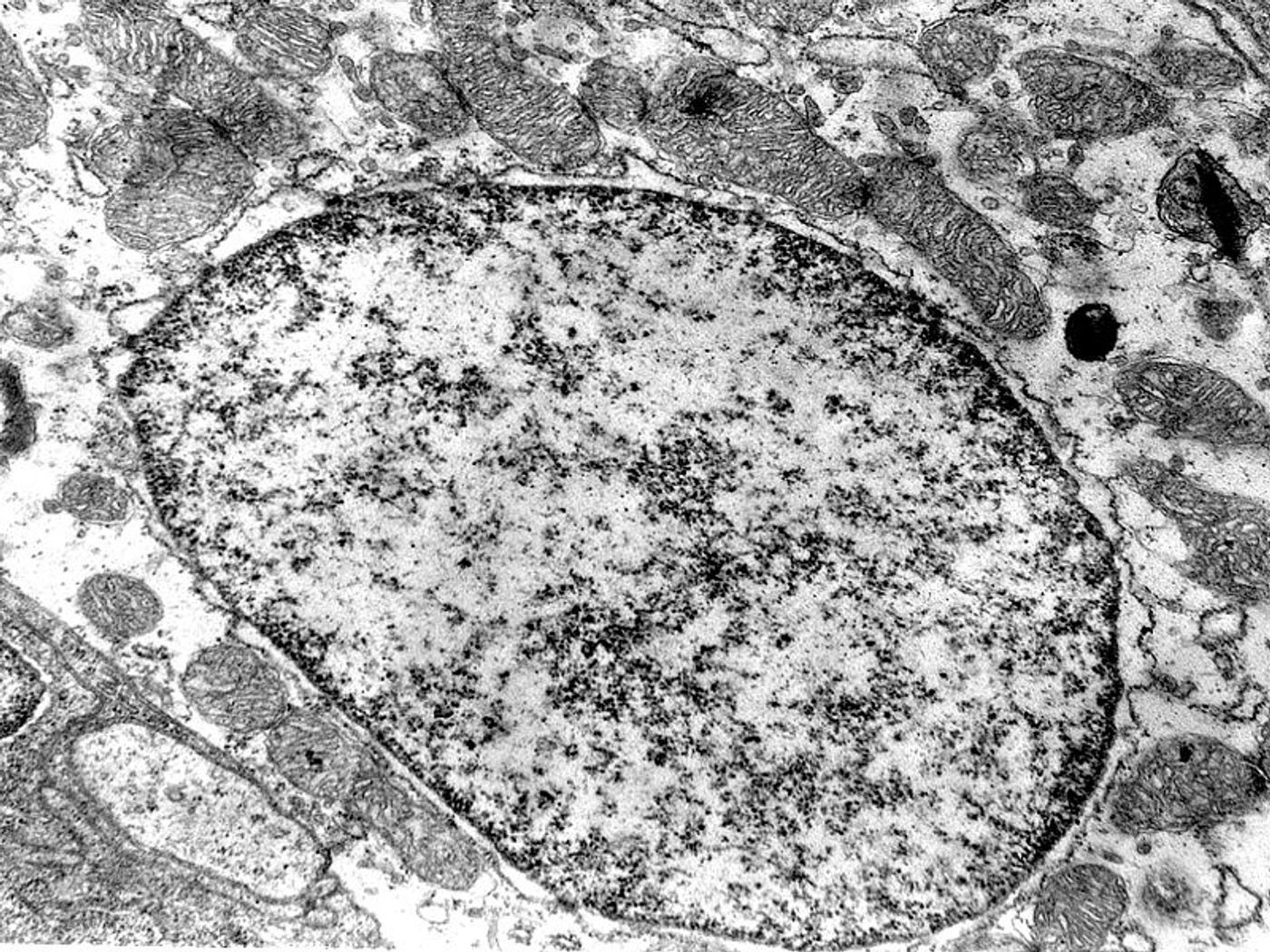
As the infamous “powerhouses of the cell,” the mitochondria are responsible for several vital roles in the cell, and thus, in the body as a whole:
In the heart, mitochondria play a particularly important role, providing energy for “optimal cardiac performance.” Thus, a disruption in mitochondrial function in cardiomyocytes will always mean a disruption in heart function.
The relationship between mitochondria and the ER
Mitochondria and the ER are physically interconnected via mitochondria-associated ER membranes (MAMs). When the connections between the two are disrupted, several diseases can develop, including Alzheimer’s and Parkinson’s, but the details of how this happens are fuzzy.
In their study, Georgia State researchers used echocardiography to monitor heart function in several situations:
The genetically deficient mice showed several issues involved in heart function, including muscle scarring, compared to control mice.
True for human hearts too?
“The formation of MAMs mediated by the mitochondrial membrane protein FUNDC1 was significantly suppressed in patients with heart failure, which provides evidence that FUNDC1 and MAMs actively participate in the development of heart failure," explained Georgia State’s Dr. Ming-Hui Zou.
The findings from the study indicate a new potential treatment approach, rejuvenating MAMs to promote both mitochondrial and cardiac function.
Going forward, Zou said, “this work has important clinical implications and provides support that restoring proper function of MAMs may be a novel target for treating heart failure."
The present study was published in the journal Circulation.
Source: Georgia State University