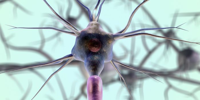
Many people have never heard of cilia, but these tiny appendages that protrude from the surface of most cells have been found to be an essential part of many cellular functions. Researchers want to learn more about them and the basis for the remarkable diversity in their roles, especially because they have been implicated in some diseases. New work reported in Nature by scientists at the Technical University of Munich (TUM) has now revealed more about how ciliary diversity arises. The findings show how a protein complex that influences the function of the cilium is activated.
Motor proteins (green dots) move along microtubules like trucks on a highway. Video by Georg Merck of TUM.
Cilia are critical to the survival of human cells, and they are also vital in many different kinds of organisms, like roundworms and flagellates. They ensure that human organs end up in the right place, and they help keep airways free of mucous. "This multifunctionality is absolutely fascinating," noted Dr. Zeynep Ökten, a biophysicist in the Physics Department of the Technical University of Munich.
The significance of the cilium has only recently come to light, but they’ve come to be known as a kind of cellular antenna that can send and receive signals. There is probably a lot more to learn about them before we know everything they do. "To date, we know very little about which biochemical processes control the various functions. This makes understanding the basic mechanisms even more important," added Ökten.
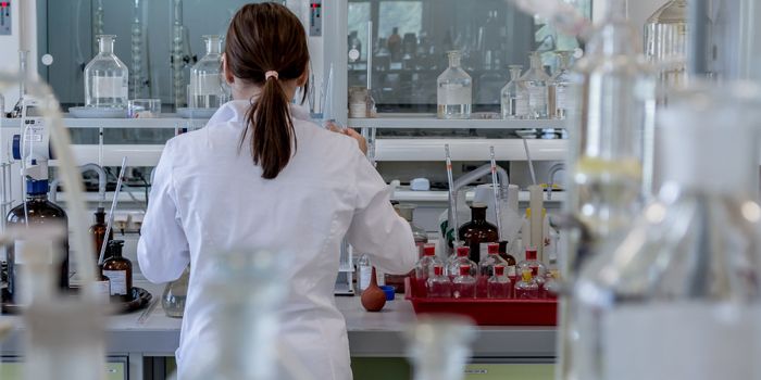
In the lab, glass plates that contain thin capillaries full of liquid are studied under a fluorescent microscope, to look for the movement of transport proteins that have been tagged with a fluorescent green marker. Those proteins are visible as dots that migrate inside the channels, as they would inside of the cilium. It’s known that these transport proteins migrate, but what moves them has been a mystery. To learn more, Zeynep Ökten and her team of researchers reconstituted the protein complex.
A common research model, the roundworm Caenorhabditis elegans is an ideal way to study the cilium. The organism uses its cilium to locate food and to detect hazards.
"Here, the classical top-down approach reaches its limits because too many building blocks are involved," explained Ökten. "To understand the intra-flagellar transport, IFT for short, we thus took the opposite approach, studying individual proteins and their interactions from the bottom up."
Related: Linking Primary Cilia and Neural Tube Defects
The researchers had to do a lot of detective work before they found the combination of proteins they were looking for. Once that minimum combination was identified, they were observed fusing into a complex that could move through capillaries.
"When we saw the images of the fluorescence microscope, we immediately knew - now we have found the parts of the puzzle that start the engine," said Ökten. "If just one of these components is missing, due to a genetic defect, for example, the machinery will fail - which, because of the cilia's importance, is reflected in a long list of serious diseases."
Knowing more about these ciliary defects that cause disease - ciliopathies - may help researchers develop disease therapeutics. Learn more about the importance of the cilium from the video below by the Salk Institute.
Sources: AAAS/Eurekalert! Via TUM, Nature