Scientists Create Neuromuscular Organoids That Contract
The muscles of the body move because of signals sent by the nervous system, which also takes in sensory information and relays it to the brain. Muscle and nerve cells meet at the neuromuscular junction. Neuromuscular disorders affect this junction or the motor neurons that are part of this system and include amyotrophic lateral sclerosis (ALS), multiple sclerosis, and muscular dystrophy. The study of these diseases and the development of therapeutics has been hindered in part by a lack of reliable cell culture models.
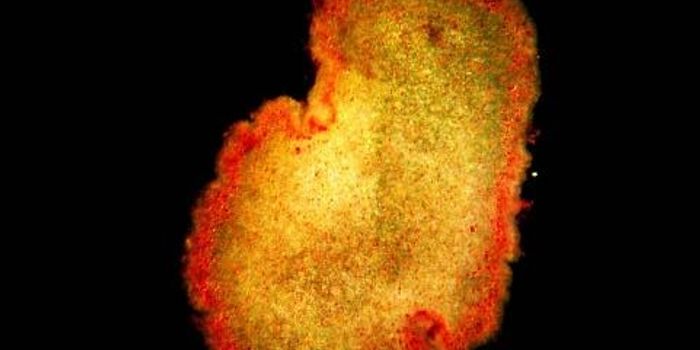
Researchers have now created a new kind of organoid, simplified versions of organs grown in the lab, which are made of cells that organize themselves into muscle and neurons, and model the neuromuscular system. In this process, the same progenitor cells give rise to motor neurons, Schwann cells, and skeletal muscle cells. Together, this group of cells sends oscillating signals through central pattern generator (CPG) circuits, which are required for functions like walking and breathing. The work, which will open up new avenues in the study of neuromuscular disease, has been reported in Cell Stem Cell.
"Our initial goal was to develop functional neuromuscular junctions, but the findings exceeded our expectations because the additional development of the CPG-like networks was an exciting but unexpected finding," said Dr. Mina Gouti, head of the Max Delbrück Center for Molecular Medicine (MDC) Stem Cell Modeling of Development and Disease Lab. "This has not been shown in a human in vitro model before, and offers entirely novel possibilities, including the study of CPG involvement in neurodegenerative diseases."
Researchers have had to grow motor neurons and muscle cells separately, then combine them to study the human neuromuscular junction, a strategy that also lacked the Schwann cells that would normally be found at the junction.
"It is very limiting if you only have an enriched system for neurons or muscles and then combine them," said the first author of the study, Jorge-Miguel Faustino Martins, a bioengineering graduate student in the Gouti lab. "It doesn't really mimic what happens in the embryo where you have both systems developing simultaneously. By combining the potential of stem cells with the powerful organoid technology, neuromuscular organoids present an exciting model to study neuromuscular diseases as well as a robust model for developmental studies where the formation of complex neuromuscular circuitry can be analyzed in real-time in [a] 3D microenvironment closer to the one present in the embryo."
The MDC scientists built on previous work and used human pluripotent stem cells to generate a specific type of axial stem cells. In 3D cell culture, these cells self-organized into the various cell types and began to mimic the CPG circuits.
"These organoids started contracting after 40 days in culture," said Gouti. "This activity was driven by the release of [the] neurotransmitter acetylcholine from the resident motor neurons in the organoid and was not due to spontaneous muscle activity seen in other systems. We were able to show that because pharmacological blocking of the acetylcholine receptors was sufficient to abolish muscle contraction."
Learn more about diseases of the neuromuscular system from the video.
Sources: AAAS/Eurekalert! via Max Delbrück Center for Molecular Medicine in the Helmholtz Association, Cell Stem Cell


