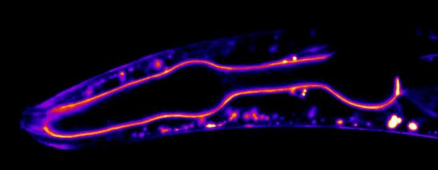
Many of the body's structures are surrounded by a matrix called the basement membrane, which acts as a kind of sheath that supports and surrounds muscles and fat and lines blood vessels and skin, for example. These membranes have been difficult to study because they are so hard to access. Scientists have now made time-lapse movies of the basement membrane by genetically modifying a nematode worm model called C. elegans so their basement membrane proteins emit fluorescence.
Basement membranes have been around since the first multicellular animals evolved on earth - more than 600 million years ago. These membranes enable organs to hold together and skin cells to stay in place. They support muscles as they stretch and compress, help filter blood in the kidneys, and send growth signals to cells during development, among other critical duties. Reporting in Developmental Cell, it's now possible to see how basement membranes develop, regenerate, and change during life. It may also provide insight into how dysfunctional basement membranes are involved in disease.
"We wouldn't be here without basement membranes," said the research leader, Duke biology professor David Sherwood.
These membranes sit deep inside the body, making them tough or impossible to see in live organisms with typical microscopy techniques. In this study, the researchers applied the CRISPR gene editor to C. elegans worms to create 29 basement membrane proteins that would glow under fluorescent light. This allowed the scientists to visualize the proteins and their movement using time-lapse microscopy.
Seeing these proteins in action, instead of having to fix tissue and proteins in place in order to study them, gives researchers a far more complete understanding of these proteins and how they function in basement membranes. Previous studies of basement membranes have relied on these fixed tissues. "As a result, they have generally been thought of as 'boring' static structures," said postdoctoral fellow Eric Hastie.
The proteins were studied and tracked in the basement membrane that lines the throat of the worm, as well as the remodeling of the membrane as it surrounded the worm's gonad; it rapidly grew to more than 90 times its original size. The movies surprised the researchers by showing that basement membranes in the worm do not typically remain in place once they're formed. Though some of the core portions are static, other proteins move within this stable architecture.
"Our findings suggest basement membranes quickly change their properties to support mechanically active tissues and they may act as highways that allow growth factors to rapidly travel," Sherwood said.
"We've just started getting to play with this tool kit," Hastie said. The researchers are hopeful that their research will offer scientists a new way to study how defects in the basement membrane are related to diseases including diabetes and muscular dystrophy.
Experienced research scientist and technical expert with authorships on over 30 peer-reviewed publications, traveler to over 70 countries, published photographer and internationally-exhibited painter, volunteer trained in disaster-response, CPR and DV counseling.