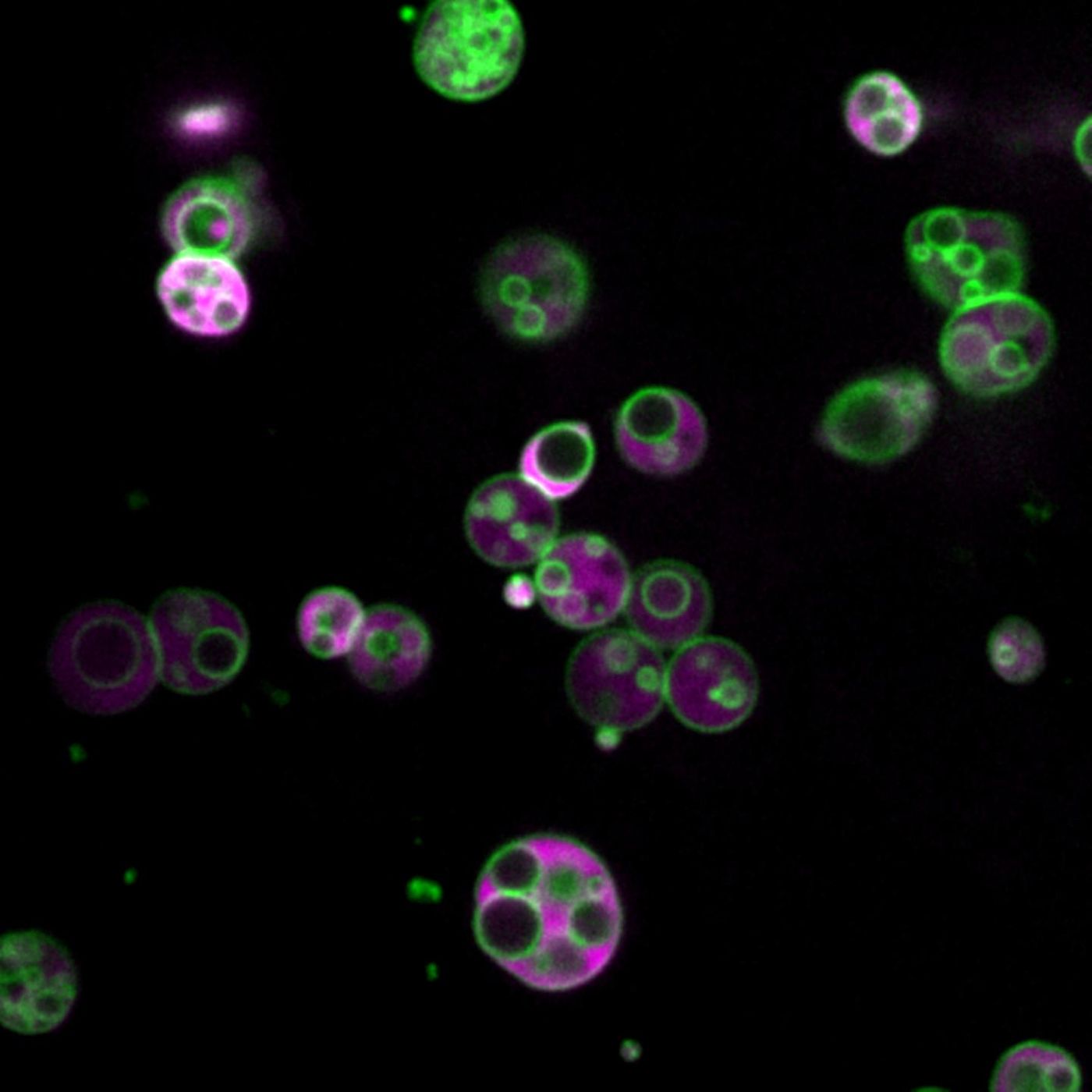
In a finding five years in the making, scientists have discovered a previously unknown compartment inside cellular organelles called peroxisomes. The findings have been reported in Nature Communications. Peroxisomes are used to break down long chain fatty acids and help degrade damaging reactive oxygen species. Dysfunctional peroxisomes have been implicated in metabolic diseases and may play a role in obesity, some cancers, neurodegeneration, or aging disorders.
The compartments were first observed in 2015 by Rice University graduate student Zachary Wright when he was using a new fluorescent tag that is added to engineered proteins. At first, his advisor thought they were looking at an artifact. Wright combed through the literature and repeated his experiments to try to learn more about what they were seeing.
"I revisited the really old literature about peroxisomes from the 60's, and saw that they had observed similar things and just didn't understand them," Wright said. "And that idea was just lost."
We know a bit about the basic structure of peroxisomes; they are like sacs that contain a granular matrix. But there is still a lot we don't know about them. The observations made in the 1960's were seen with transmission electron microscopes, a technique that was used less often after confocal microscopy was developed.
"It's just much easier than electron microscopy," said study co-author Bonnie Bartel, Wright's Ph.D. advisor. "The whole field started doing confocal microscopy. And in the early days of confocal microscopy, the proteins just weren't that bright."
Wright also used confocal microscopy, but his fluorescent reporters were bright enough to resolve small details, illuminating the compartments. The peroxisomes he was looking at were also from Arabidopsis seedlings; during germination, these cells have peroxisomes that are up to 100 times bigger than those in mammalian cells.
"One reason this was forgotten is because peroxisomes in yeast and mammalian cells are smaller than the resolution of light. With fluorescence microscopy, you could only ever see a dot. That's just the limit that light can do," Wright said. "Bright fluorescent proteins, in combination with much bigger peroxisomes in Arabidopsis, made it extremely apparent, and much easier, to see this."
"This is, without a doubt, the most unexpected thing our lab has ever discovered," Bartel noted. "This requires us to rethink everything we thought we knew about peroxisomes."
Since peroxisomes are found in all kinds of cells, including plant cells and human cells, the researchers suggested that these observations may be true of peroxisomes in general.
"Peroxisomes are a basic organelle that has been with eukaryotes for a very long time, and there have been observations across eukaryotes, often in particular mutants, where the peroxisomes are either bigger or less packed with proteins, and thus easier to visualize," Bartel said. But larger peroxisomes have been associated with mutations, so the observations were mostly disregarded.
More work will be needed to learn more about the function of these compartments, and whether they are relevant to human health and disease.
Bartel said this work will help us gain insight into peroxisomal disorders, which are described in the video.
"This work could give us a way to understand some of the symptoms, and potentially to investigate the biochemistry that's causing them," she said.
Experienced research scientist and technical expert with authorships on over 30 peer-reviewed publications, traveler to over 70 countries, published photographer and internationally-exhibited painter, volunteer trained in disaster-response, CPR and DV counseling.