Microvillus inclusion disease (MVID) is a cause of chronic watery diarrhea that is attributed to a lack of nutrient absorption in the gastrointestinal tract in newborns. The disease can lead to malnutrition and dehydration which can be life-threatening. Other complications include difficulty gaining weight, failure to thrive, liver and kidney problems and osteoporosis. Some individuals will develop cholestasis, or reduced ability to produce bile, which can also lead to cirrhosis of the liver. Typically MVID is a lifelong illness that requires nutritional support and most children suffering from MVID do not survive passed childhood.
The prevalence of MVID is unknown, however is has been found to occur worldwide. It is inherited through an autosomal recessive pattern indicating that both copies of the gene that are inherited have a mutation. This means that both parents of an individual with MVID are each asymptomatic carriers of the mutated gene which causes MVID.
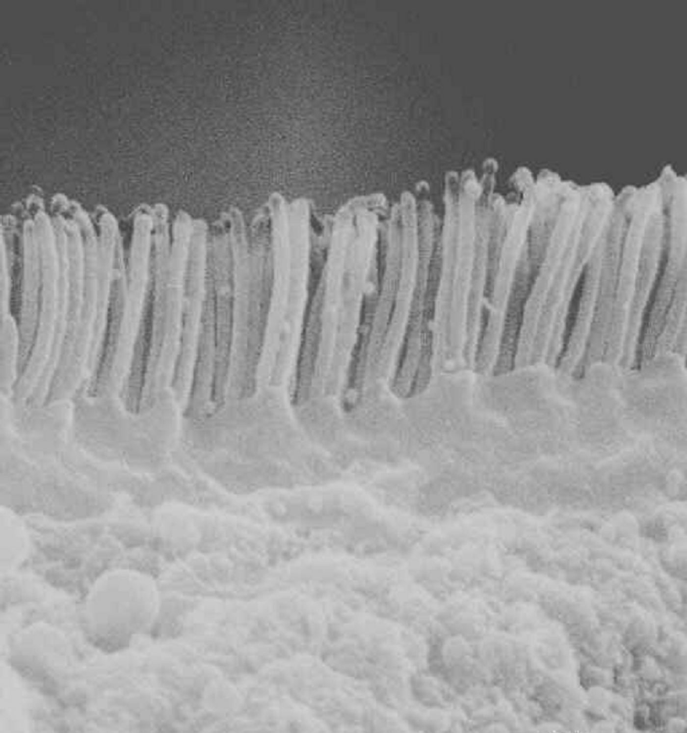
The mutation which causes MVID is thought to affect the MYO5B (myosin-Vb) gene. The protein myosin-Vb belongs to a group of proteins with similar structures known as myosins. Myosins are involved in cell movement and transport of materials between cells. They also help determine the position of components within cells, which is also known as cell polarity. In individuals suffering from MVID, it is hypothesized that there is a depletion of apical microvilli and the formation of microvillus inclusions inside the cells, suggesting the loss of polarity.
A research team from France sought to investigate the hypothesis of polarity loss by examining the location of essential polarity determinants in five individuals suffering from MVID. They collected small bowel biopsies from patients suffering from MVID as well as individuals not suffering from MVID (control group). Immunohistochemistry staining was performed to detect antigens in affected tissues. The tissues were also examined by electron microscopy to compare the cell surface topography of affected and normal tissues. Lastly, colorectal adenocarcinoma cells (Caco-2) with the MYO5B gene silenced using interfering RNA were used to quantification of cysts central and/or basal location of polarity determinants.
Researchers found that specific polarity determinants and structural proteins were lost from the apical membrane in MVID affected tissues. The polarity determinants and structural proteins were found to be accumulated in the cytoplasm or basal side of enterocytes. This indicated the inversion of cell polarity in tissues from individuals suffering from MVID. Formation of microvilli like structures on the basal side of enterocytes was confirmed in these tissues by electron microscopy. Their experiments using Caco-2 cell lines with a silenced MYO5B gene showed that there is a direct link between the loss of the gene and mislocation of apical proteins.
The results of this study confirm that silencing of the MYO5B gene leads to polarity inversion in enterocytes. This loss of polarity was specific for tissues from individuals diagnosed with MVID. Specific polarity determinants, including Cdc42 which is a protein coding gene involved in cell division, was identified as a potential biomarker for the disease.
Sources:
Biology of the Cell;
National Institutes of Health