Using powerful microscopy techniques, researchers have learned more about autophagy, a waste recycling process employed by cells, and
published their results in Nature Communications. The scientists used live imaging to actually see the cells encapsulate and recycle stuff at the molecular level.
Autophagy [aw-
tof-uh-jee] is a mechanism in which cellular contents are gathered, packaged and turned over into new molecules and structures. As a fundamental part of cellular function, any errors in this process can result in disease. Such disorders in which cellular debris is not cleared away can be related to aging, as in Alzheimer’s, cancer or rheumatoid arthritis. For more information about autophagy, check out the video below.
In this work, the investigators honed in on a characterizing a structure that is only seen at the very beginning of the autophagy process and is transient. It functions to envelop material meant for degradation and gives rise to the autophagosome, a vesicle or small sac within the cell that ultimately deliver its contents to the lysosome for breakdown.
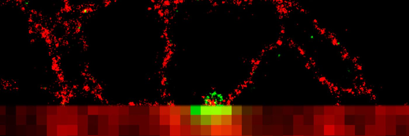
The short-lived nature of the structure presented challenges to the researchers. As such, a new approach was developed that harnessed and combined two techniques. Investigators working at the Babraham Institute used live imaging microscopy followed by dStorm (direct Stochastic Optical Reconstruction Microscopy). A short demonstrative video is seen below, showing what individual labelled molecules can look like when using the technique.
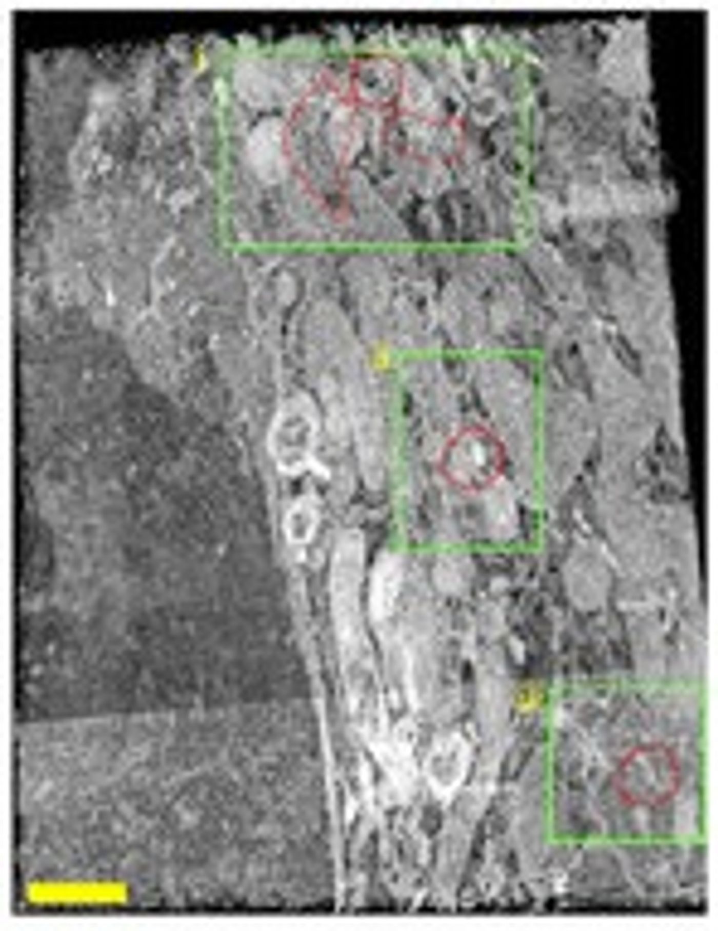
The second method, utilized by scientists collaborating at the Francis Crick Institute in London and the Zeiss Microscopy Labs in Munich, is called FIB-SEM (Focused Ion Beam Scanning Electron Microscopy). This video shows what Focused Ion Beam Scanning Electron Microscopy image looks like as the process is performed.
The combined power of the methods enabled researchers to show how the first structure forms when autophagy initiates, and elucidate the various protein and membrane interactions that lead it to develop into a mature autophagosome.
The group leader in the Signaling research program at the Babraham Institute and senior author of the new work, Dr Nicholas Ktistakis, comments, "By combining live imaging with cutting-edge super resolution microscopy techniques, we have been able to characterize the site of autophagy initiation and observe the physical and functional interactions between the proteins involved in autophagy. This has uncovered a new level of detail of the earliest stages of autophagy and provides a general protocol for this type of analysis in other areas of cell biology.
"Knowing more about this process increases our ability to find ways to manipulate or boost it for future therapeutic benefit," he concludes.
Sources:
Nature Communications,
AAAS/Eurekalert! via
Babraham Institute