In cells, not all genes are active, or being used by the cellular machinery at any given time. The right genes have to be expressed at the right times in order to keep the physiology of organisms maintained and functioning properly. Researchers often study gene expression in order to better understand organisms in development, and the healthy and diseased states of mature tissues, for example. There are a variety of different ways to assay gene expression, and of course they all have their advantages and disadvantages. Scientists at Caltech have now invented a new way to get a look at gene expression inside the body, published in Nature Communicatons. Visualizing gene expression usually involves using transgenic animals or studying cells in culture, and previous atempts to use MRI to visualize gene expression have needed improvements.
Led by Mikhail Shapiro, an Assistant Professor of Chemical Engineering and Heritage Medical Research Institute Investigator, investigators have created a technique that fuses the signals detected by magnetic resonance imaging (MRI) to gene expression. This could be a way to perform a non-invasive biopsy; doctors could look at gene expression and determine if tumors were present in a patient’s tissues without having to perform surgery.
The MRI method works because hydrogen atoms contained in the human body, within water molecules and fat, can be excited when exposed to a magnetic field, which just passes right through tissue. The excited hydrogen atoms emit signals that are subsequently used to make images of the internal structures of the body, distinguishable based on location and environment. It’s a common technique for producing a snapshot of internal physiology, but has not been used for imaging at the cellular level.
"We thought that if we could link signals from water molecules to the expression of genes of interest, we could change the way the cell looks under MRI," explained co-lead author of the work, Arnab Mukherjee, a postdoctoral scholar in chemical engineering at Caltech.
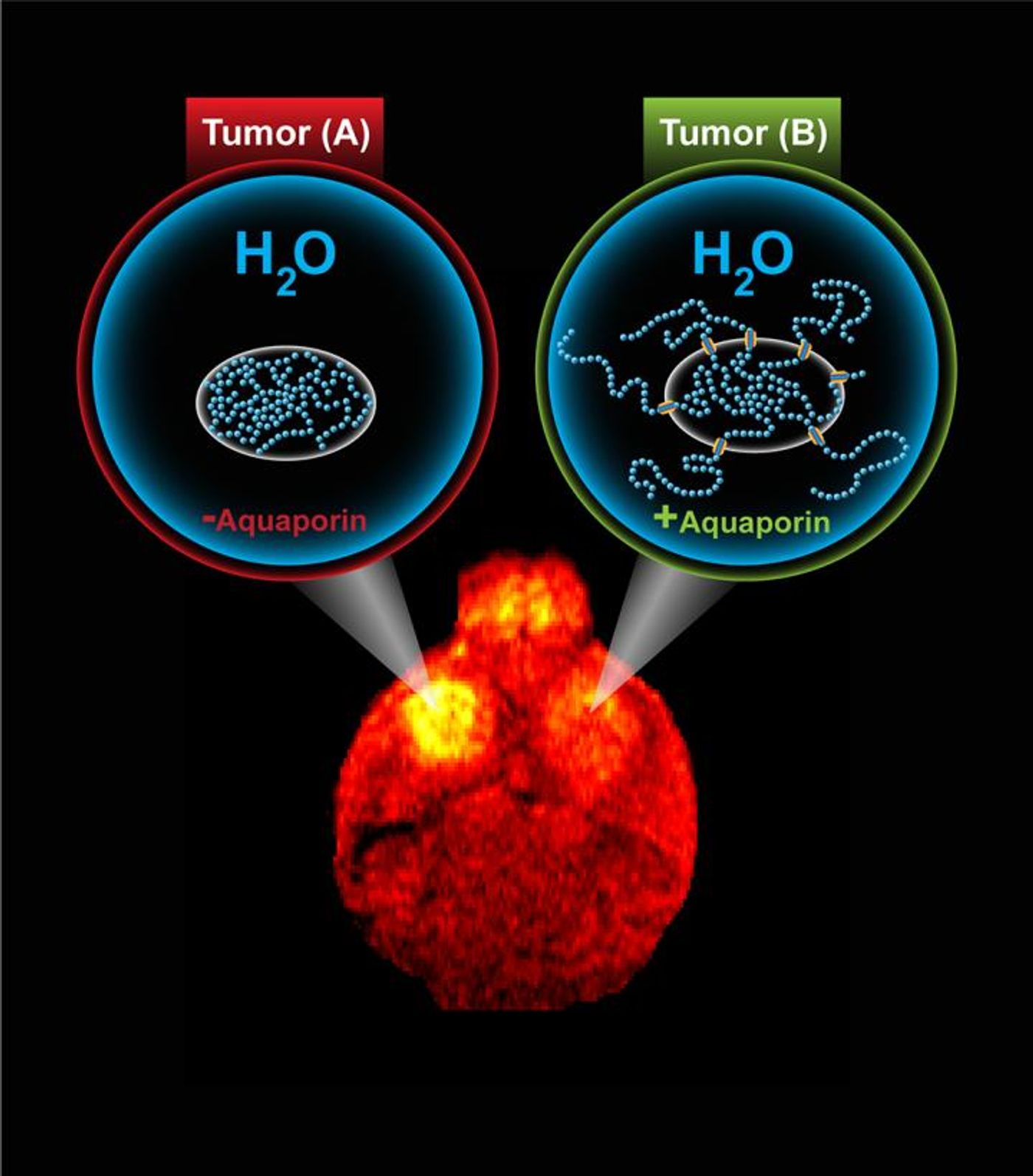
For their research, the team used a molecule called aquaporin, which acts as a channel for water molecules to enter and exit the membrane that surrounds cells. Increasing the amount of aquaporin on a cell made it stand out when the researchers used a procedure called diffusion-weighted imaging to acquire their MRIs. That made aquaporin good to use as a reporter – something that will indicate experimentally that a gene is active. When a gene of interest is linked to the aquaporin reporter, in cells where the gene is being expressed, the cell will appear darker using diffusion-weighted imaging.
As a proof-of-concept, the researchers monitored gene expression within a brain tumor in a mouse. The tumor was implanted, and the mouse was given a drug that triggered the expression of the aquaporin reporter gene in the tumor. The tumor then looked dark in MRI scans.
"Overexpression of aquaporin has no negative impact on cells because it is exclusive to water and simply allows the molecules to go back and forth across the cell membrane," Shapiro said. The amount of water in a cell stays constant; under typical conditions the number of water molecules that enter and exit a cell expressing aquaporin are equal. "Aquaporin is a very convenient way to genetically change the way that cells look under MRI."
Shapiro thinks that while this work used mice for the model, it could be used in the clinic one day. Other techniques that have used MRI reported genes have been limited because they’ve needed metals not always present in tissue. Aquaporin, however, is naturally occurring and should not elicit a reaction from the immune system.
"An effective reporter gene for MRI is a 'holy grail' in biomedical imaging because it would allow cellular function to be observed non-invasively," said Shapiro. "Aquaporins are a new way to think about this problem. It is remarkable that simply allowing water molecules to more easily get into and out of cells in a tissue gives us the ability to remotely see those cells in the middle of the body."
If you would like to know more about the physics behind MRIs used on patients, watch the above video. The video below has more information about aquaporins.
Sources: AAAS/Eurekalert! via Caltech, Nature Communications