Proton pumps are specialized proteins that reside in the envelope that surrounds a cell, the cell membrane, and the pumps can transport protons across that membrane in one direction. The unidirectional proton movement can create a pH and charge gradient capable of acting as a power source for cellular functions. Researchers have now used powerful tools to learn more about the proton pumps by studying one in particular, bacteriorhodopsin.
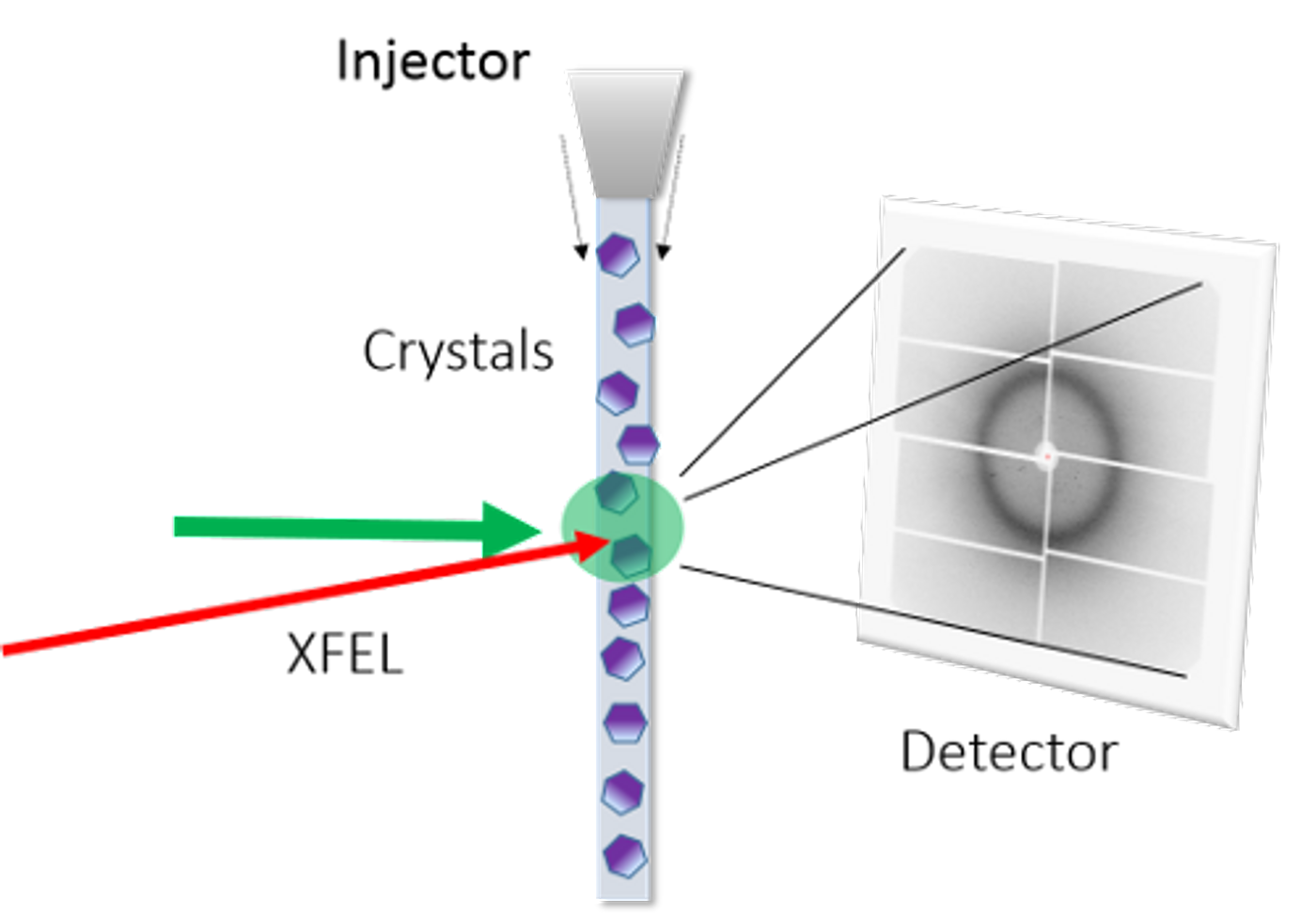
Working with collaborators, investigators at the RIKEN SPring-8 Center utilized SACLA, an X-ray Free Electron Laser. X-rays are capable of imaging at the atomic level because it is a light source with a much shorter wavelength than visible light, and SACLA is over a million times brighter than typical facilities. Passing samples through an x-ray beam vaporizes it, however, because of the strength of the beam. As such, images have to be captured rapidly, in the instant before they are destroyed.
In this work, published in Science, researchers applied time-resolved femtosecond crystallography, in which thousands of images of bacteriorhodopsin are obtained after light strikes. This all enabled the researchers to understand the ability of the pump to push protons into a more positively charged environment outside of the cell compared to the inside, which makes a sort of battery that powers chemical reactions in the cell.
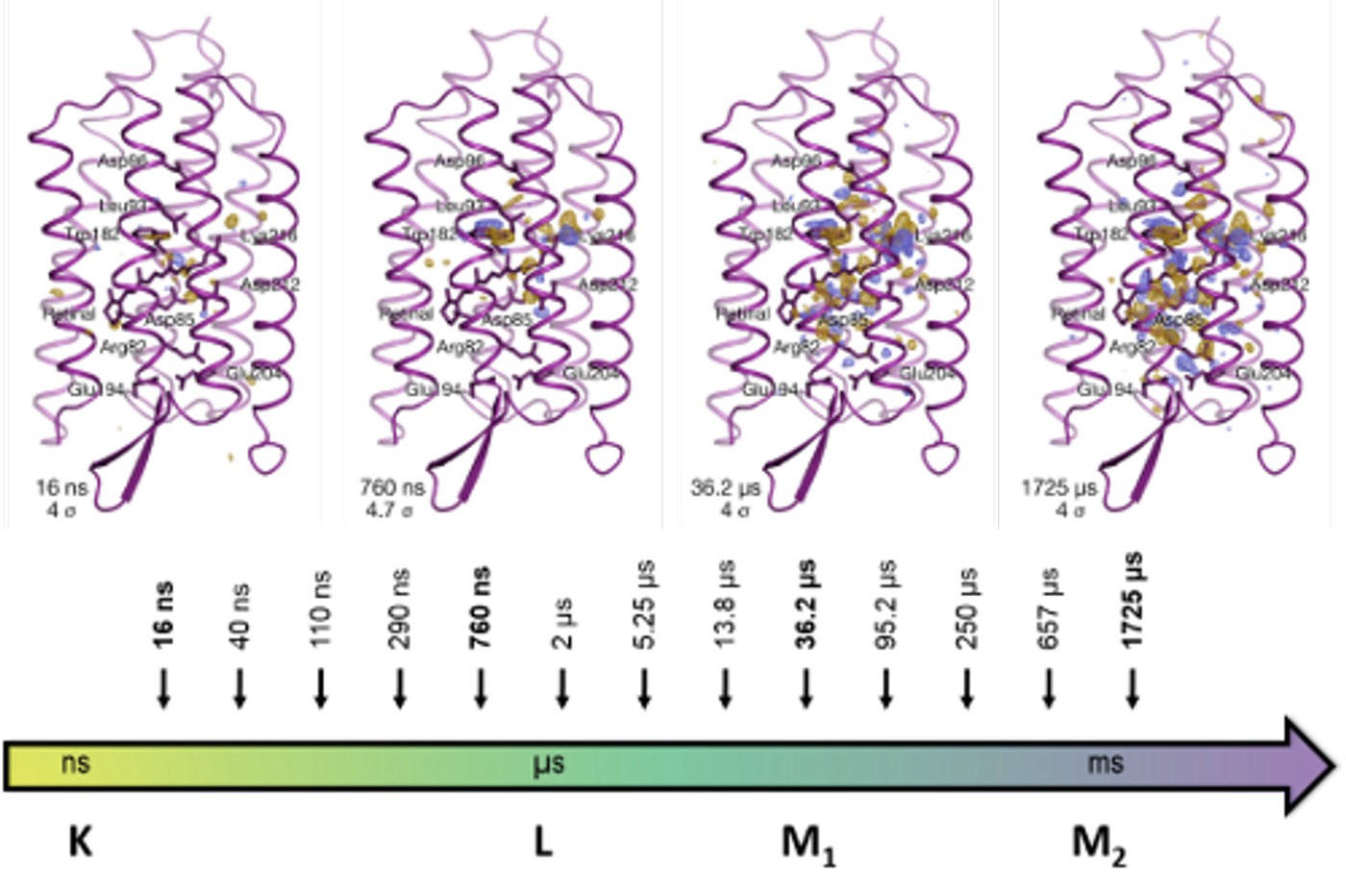
"With this work, we were able to shed light on the mechanism of proton transfer and bring closure to a long-standing debate regarding the mechanism. The photoexcitation leads to a change of conformation in retinal, the key part of bacteriorhodopsin that absorbs the light. This in turns leads to the displacement of amino acid residues above the retinal towards the cytoplasm, and a transient water appears in that space to assist the primary proton transfer. Following this, a delicate molecular cascade prevents the proton from moving backward, and it is pushed up outside of the cell,” commented Eriko Nango, the first author of the study.
Many of the details of the proton pump mechanism were known, but questioned due to suspicions that the radiation used to study it could be inducing alterations. This new technique allowed the scientists to characterize it conclusively. The video below illustrates some of those details - although this is not the video from the study.
“New techniques using powerful x-ray lasers are giving us insights into precisely how processes such as proton pumping take place in actual biological systems,” said So Iwata, the leader of the study. “This will give us much greater understanding of these actions, and ultimately lead to insights that will allow us to manipulate them more effectively."
You can learn more about bacteriorhodopsin in the following video.
Sources: AAAS/Eurekalert! via RIKEN, Biophysical Journal, Science