The nervous system is incredibly complex and begins developing very early on in life, then goes on to be the last one finished. New work has described a pattern of organization among cells that create the spinal cord, finally revealing it in precise detail. This new data could aid the field of regenerative medicine; knowing how cells form structures will help inform strategies to get them to form once again if they are damaged.
Cell patterning is critical throughout development; the right cells have to arrive in the proper location to ensure an organism is formed correctly. When it comes to the nervous system, nerve cells have to organize into circuits that transmit the right information at the correct time to the right places, or else the body and brain will not be able to control the muscles and move properly. This work has now shed light on how this organization happens, something that was not well understood before now.
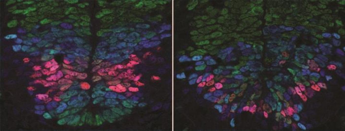
Reporting their findings in Science, scientists collaborating at the Francis Crick Institute, the Institute of Science and Technology (Austria) and Ecole Polytechnique Fédérale de Lausanne (Switzerland) learned that cells which eventually become nerve cells in the developing embryo of a mouse utilize two different signals that emanate from opposite sides of the spinal cord, enabling them to accurately sense their positions. Based on this geography, these cells then become the nerve cell type they should be.
The team included biologists, engineers and physicists who all worked to determine that the levels of the two signals that come from opposing sids of the body - back and belly - can exert an influence on the genes that are expressed in developing nerve cells. That gene expression which was happening in early development helped the cells become the right nerve cell type for where they are located in the spinal cord.
"We've made an important step in understanding how the diverse cell types in the spinal cord of a developing embryo are organized in a precise spatial pattern. The quantitative measurements and new experimental techniques we used, as well as the combined effort of biologists, physicists and engineers were key. This allowed us to gain new insight into the exquisite accuracy of embryonic development and revealed that cells have remarkable ability of to orchestrate precise tissue development," explained Anna Kicheva, the Group Leader at IST Austria.
"We have shed light on the long-standing question of how developing tissues produce the right cells in the right place in the right numbers," said James Briscoe, the Group Leader at the Francis Crick Institute. "It's likely that similar strategies are used in other developing tissues and our findings might be relevant to these cases. In the long run this will help inform the use of stem cells in approaches such as tissue engineering and regenerative medicine. However, there is still much more to learn and we need to continue developing these interdisciplinary collaborations to further our biological understanding."
Learn more about how cell patterning works in the developing embryo from the video, which is a talk by Eric Wieschaus of Princeton.
Sources: AAAS/Eurekalert! via Crick Institute, Science