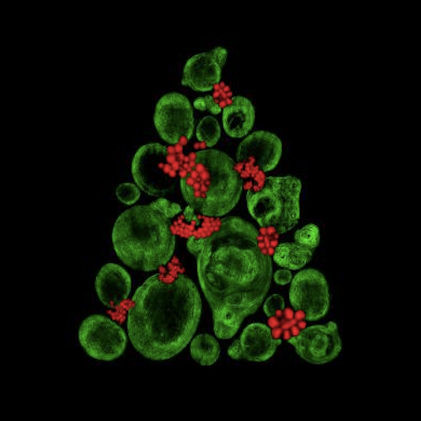
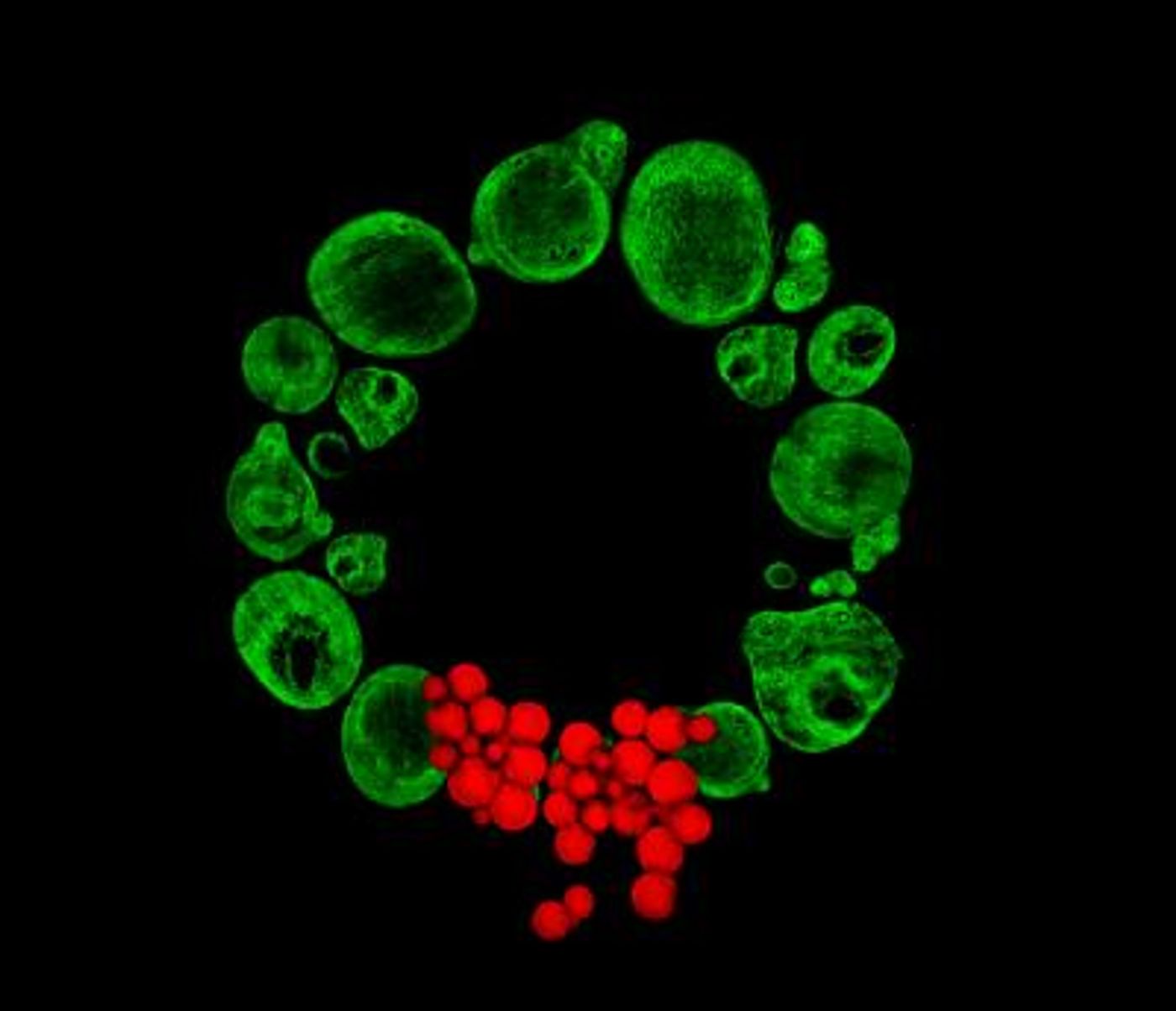
The seasonal theme of these gorgeous images was generated using a new laser-based imaging technique; they were created by University of Southampton postgraduate researcher Catarina Moura. Seen in green are collagen cells, and in red, fat cells, both of which were taken from human bone marrow. This is part of a project aiming to develop new tools to monitor the development of cells used in regenerative medicine.
In this research, the investigators want to use human stem cells to grow cartilage. Moura is working to engineer a way to image human stem cells during skeletal regeneration in a non-invasive and label-free way.
"I've chosen green for the collagen fibers in my images because when you use the labelling technique typically you use a stain that fluoresces as green and, because a scientist would usually relate to that green color when looking at labelled collagen fibers, I decided to use the same color and create something more festive at the same time," noted Moura.
"I'm working with Professor Richard Oreffo and Dr Rahul Tare from the University's Centre for Human Development, Stem Cells and Regeneration who are trying to create and grow cartilage in the lab using a patients' own (autologous) stem cells to then be implanted back into the patient if they have a cartilage defect," she explained. "My part of the project is to develop and use techniques to make it easier monitor the development of the cells into cartilage in real time which is important to knowing if and when you can use it for the patient. If it's successful, you can use the same cartilage to create the new tissue so it's very important for us to get the monitoring right." Oreffo is featured in the video at the bottom of the article.
Typical tools use stains or fluorescent molecules to label the structures inside of cells. Moura, with the help of her Ph.D. supervisor, Southampton's Sumeet Mahajan, Professor in Molecular Biophotonics & Imaging in Chemistry & Institute for Life Sciences (IfLS), is using ultra-fast lasers to get the same result in less invasive ways.
"Traditional techniques to detect whether the cartilage is developing can be disruptive and, in many cases, destructive," Catarina explained. “Our process has not been used before. What we are trying to do is introduce to biology techniques normally used in chemistry or physics, using inherent chemical or structural properties of the human stem cells. Currently, for validation we still need to do the standard exercises alongside our new techniques to be able to compare the two sets of results and, of course, using ultra-fast lasers we need to ensure that everything is optimized before it can go to the clinic, especially the exposure time.
"The massive advantage with our stain-less laser-based imaging approaches is that you can use the stem cell sample without having to interrupt the developmental process in real time, you don't need to perform any cell disruption and there is no photobleaching (fading) which is fairly common with fluorescent material," Catarina continued. "Just put the bioengineered cartilage under the microscope and you have the image."
"Crucially, unlike current standard staining-based methods the stain-less imaging approach is translatable to the clinic as the stem cells are not harmed or disrupted in any way. Hence, the technology can be used to objectively assess development and screen stem cells to be absolutely sure before using them for therapy,” said Professor Richard Oreffo.
"This work perfectly exemplifies highly exciting cross-faculty interdisciplinary research that is pushing boundaries to achieve high impact. Ph.D. funding by the University's Institute for Life Sciences for Catarina kick-started the collaboration between Richard and Rahul at the Institute for Developmental Sciences and us, which otherwise might have been difficult, that has led to exciting results, some stunning images, and insight that has the potential to change people's lives using stem cell therapy,” concluded Mahajan.
Sources: AAAS/Eurekalert! Via University of Southampton