Scientists at the University of Southern California created a freely accessible online kidney atlas. They are hopeful that scientists will use it to engineer new therapeutics and techniques for treating kidney disease, which impacts about 30 million Americans. This research can also aid in the development of miniature kidneys that are more realistic and will better research models. The data has been reported across three new studies published in the Journal of the American Society of Nephrology.
"Stem-cell-based technologies hold great promise for developing kidney replacement and regeneration therapies," said the first author of the studies, Nils Lindstrom, a research associate in Stem Cell Biology and Regenerative Medicine at the Keck School of Medicine of USC. "Getting there requires detailed knowledge of how kidneys normally form so the process can be replicated in cell cultures in the lab. Our data will help us and other scientists improve current techniques to make better tiny functional kidneys."
For four years, stem cell investigators at USC and computer scientists at USC Viterbi School of Engineering have outlined the differences and similarities between the molecular and cellular characteristics of human and mouse kidney formation. They want to improve treatments for kidney disease.
The lab of senior author Andrew McMahon, Professor of Stem Cell Biology and Regenerative Medicine and Biological Sciences at the Keck School of Medicine, has been focused on the kidneys for decades.
"Our research bridges a critical gap between animal models and human applications," McMahon said. "The data we collected and analyzed creates a knowledge base that will accelerate stem-cell-based technologies to produce mini-kidneys that accurately represent human kidneys for biomedical screening and replacement therapies."
The first high-resolution atlas for human kidney development is available here: http://www.gudmap.org.
In this study, the scientists compared 26 genes that are critical for human kidneys to their mouse counterparts. Of those genes, which are anchor genes necessary for development, only eleven percent were expressed in comparable ways in both organisms: SLC22A6, ENTPD5, and UMOD. "If the goal is to treat human kidney disease, clearly, it's better to focus on genes that are also active in human kidneys," McMahon explained.
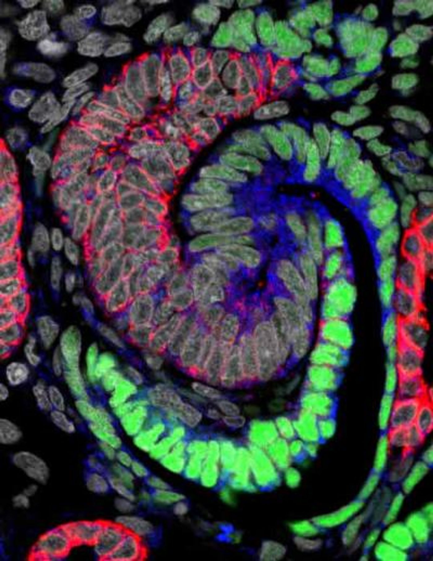
Kidney development begins with a population of 'progenitor cells' (green), which are similar to stem cells. Some progenitor cells (red) stream out and aggregate into a ball, the renal vesicle (gold). As each renal vesicle grows, it radically morphs into a series of shapes -- can you spot the two S-shaped bodies (green-orange-pink structures) -- and finally forms a nephron. Each human kidney contains one million mature nephrons, which form an expansive tubular network (white) that filters the blood, ensuring a constant environment for all of our body's functions. / Credit: Video courtesy of Nils Lindstorm, Andy McMahon, Seth Ruffins and the Microscopy Core Facility at the Eli and Edythe Broad Center for Regenerative Medicine and Stem Cell Research at the Keck School of Medicine of USC
A variety of common health conditions like diabetes, high blood pressure, and recurrent kidney infections can lead to kidney disease, which can also be caused by genetic mutations. Patients have to go on dialysis while waiting for a kidney transplant, and the median wait time is over three years. The scientists took a multi-pronged approach to tackle these health problems.
Study co-author Carl Kesselman, Dean's Professor of Industrial and Systems Engineering at USC Viterbi, led a team that created software to automate some aspects of data collection. The program, DERIVA (Discovery Environment for Relational Information and Versioned Assets), uses microscopes to gather images and organize them with other information into photo albums that can easily be viewed and shared.
"If you think of data as the modern version of a book, we gave the researcher tools to write the book, made the library where the book is stored and created a catalog system so others can find the book and check it out," said Kesselman, a principal investigator at the USC Michelson Center for Convergent Bioscience.
"It's the difference between taking a photo with film or taking it with your smartphone and using the metadata that is automatically generated to organize your photos," Kesselman explained. "At some point, you have so many photos that they become unmanageable. You can't find the one you want. DERIVA automatically catalogs your data so you can analyze massive amounts of data with Zen-like calm and share them with all of your friends throughout the world."
Soucres: AAAS/Eurekalert! via USC, Journal of the American Society of Nephrology