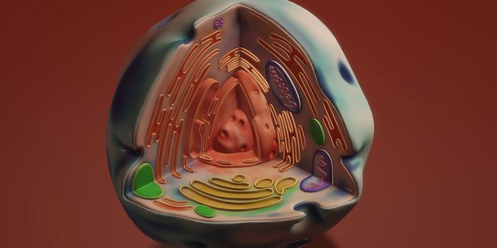
Every cell in our body is encased by a membrane; there are a variety of ways that molecules can move across that enclosure, and now researchers have learned more about that movement. With an imaging technique they created for this study, the research team of Professor Akihiro Kusumi and colleagues used fluorescent markers to watch proteins dance across the membrane.
"The proteins in the cell membrane undergo elegantly coordinated dances to relay messages between the cell and its environment,” said Kusumi, leader of the Membrane Cooperativity Unit at the Okinawa Institute of Science and Technology Graduate University (OIST),
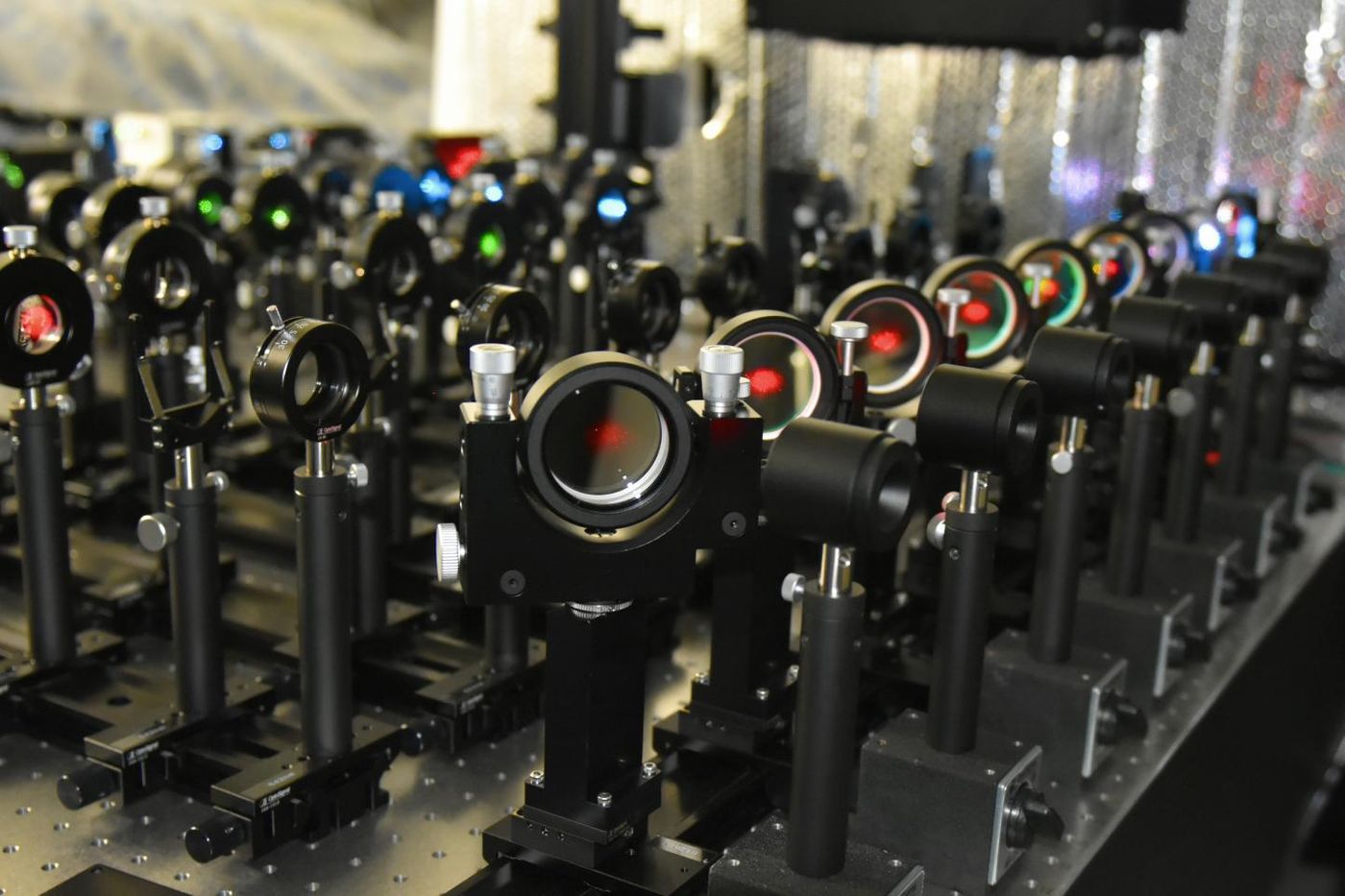
The new imaging method can capture events in live cells and is termed live-cell single fluorescent-molecule imaging (SFMI). With fluorescent microscopes, investigators can visualize movement in the membrane. There are challenges in fluorescent microscopy, however. Exposing fluorescent molecules to the intense light of a microscope can cause photobleaching, where the glow is lost from the markers. Scientists were limited to about ten seconds of imaging.
"It was like randomly taking many ten-second long clips, and trying to connect them in the correct order to produce a movie lasting for five minutes," said Mr. Taka-aki Tsunoyama, a researcher from the Membrane Cooperativity Unit at OIST.
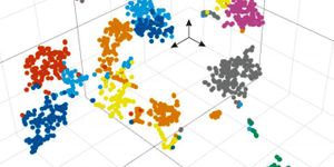
This movie shows many integrin molecules moving, and it's fast-forwarded 6x. The focal adhesion is blue; the green spots represent single integrin molecules. The yellow lines show the trajectory of the integrin molecule. Temporary immobilization is shown in red.
Tsunoyama, Kusumi, and colleagues have reported in Nature Chemical Biology that they can reduce photobleaching with a combination of chemicals and molecular oxygen. By placing the cells in a low-oxygen environment and adding the chemicals Trolox and Trolox quinone, photobleaching was significantly reduced without affecting cell viability. Now the markers can be seen for up to 400 seconds.
"Our method improves the observation time of fluorescent molecules by forty-fold," said Kusumi.
The extended time opened new doors for the scientists. They were able to see areas of the membrane called focal adhesions, which allow the cell to migrate. "These are effectively the feet of the cell," explained Kusumi.
Within the focal adhesion are groups of proteins called integrins; they connect the internal framework of a cell to its extracellular matrix. By watching for a longer time, the researchers saw that integrins move repeatedly and halt within and between feet.
The integrins move around until they find a new point of attachment, binding when one is found. After stability is restored, the integrin pulls the cell forward.
"With our method, we can now follow the movement of each individual molecular dancer for sufficiently long periods of time to understand the cellular context," added Kusumi.
Understanding focal adhesion points will lead to a better understanding of cancer metastasis, and may help stop it, said Kusumi.
Sources: AAAS via OIST, Nature Chemical Biology