Tissue bioengineering is rapidly expanding as the demand for lab-grown tissues grows steadily due to shortages in donated material for transplantation. Three-dimensional (3D) printing technology holds immense potential for engineering tissues in the lab. And most recently, researchers at Wake Forest Baptist Medical Center
demonstrated the full utility and feasibility of this technology in printing complex human-scale tissues that model the structural integrity of their biological counterparts.
In 3D printing, various materials are layered and formed into multi-dimensional structures. When applied to tissue engineering, the idea is to use biocompatible materials and actual live cells as the “ink” to be printed into tissues.
The printer up for this job is the Integrated Tissue and Organ Printing System (ITOP), a device which took an impressive 10 years to develop by scientists at the Wake Forest Institute for Regenerative Medicine (WFIRM). As “ink,” this machine uses bio-degradable and plastic-like materials to create the 3D shape of the structure, which also contain live cells in a water-based gel matrix. This system can produce biological tissues at the right size and structural integrity for implantation into the body. However, the tissue must also be strong enough to survive and integrate into the body after implantation.
Led by Anthony Atala, director of WFIRM, the research team optimized the ITOP printer to produce healthier and more resilient tissues. They incorporated micro-channels into the tissue constructs, which allowed diffusion of nutrients to the printed cells and conferred longer survival times. This new property combined with a modified “ink” that holds the cells ensured the bioprinted tissues survived long enough to be incorporated into the body.
"Our results indicate that the bio-ink combination we used, combined with the micro-channels, provides the right environment to keep the cells alive and to support cell and tissue growth," said Atala.
In order to tailor print tissues for patients, the team also incorporated clinical imaging data from CT and MRI scans as part of ITOP’s input. This allows the 3D printer to size-match tissues for each patient.
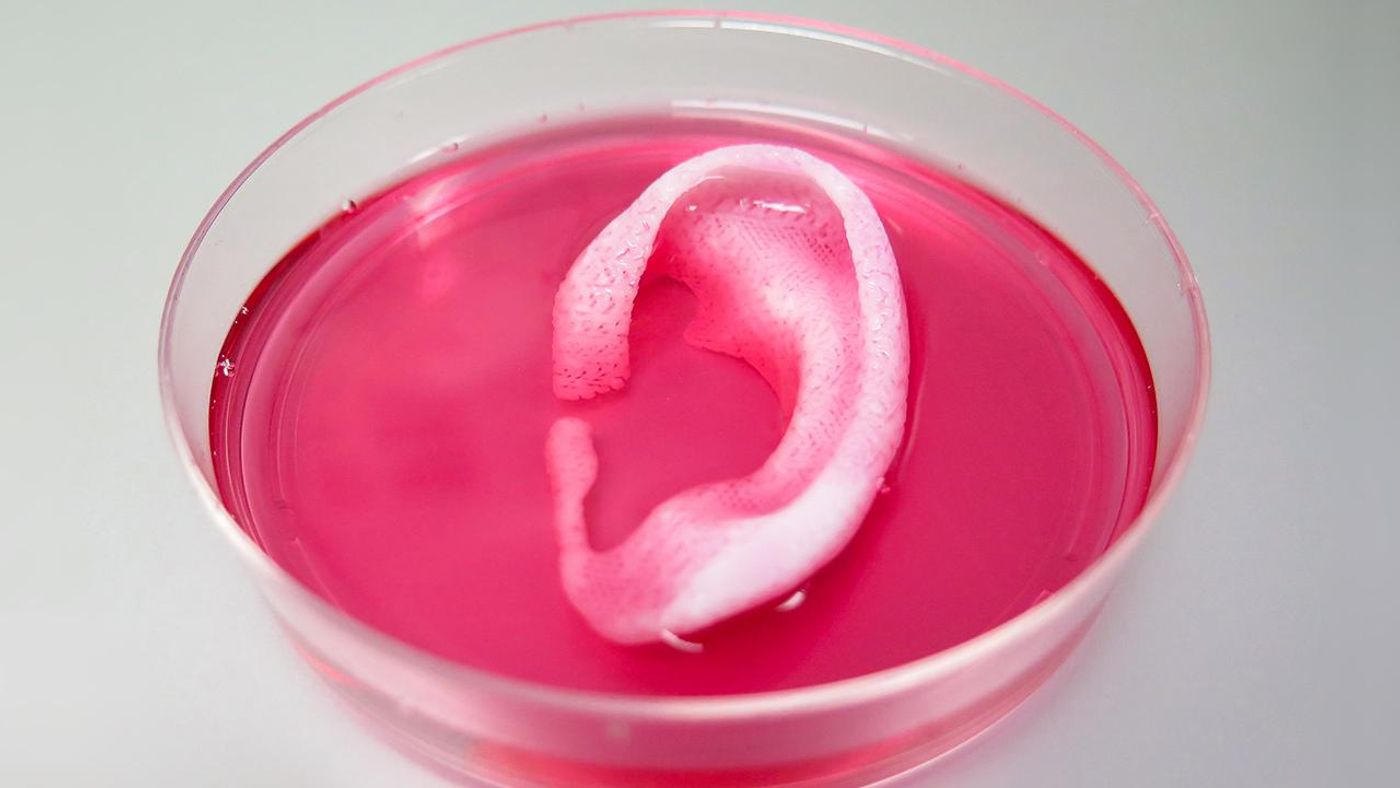
With the modifications, ITOP printed a replica human-sized ear based on a CT image. The ear cartilage structure was able to mature after implantation under the skin of a mouse. After 2 months of implantation, the artificial printed ear not only maintained structural integrity, but also developed its own network of blood vessels.
Moving to human muscles, a more organized type of soft tissue, the team used mouse myoblasts to make a 3D muscle construct. They noted that the printed structure had muscle fiber-like bundles, and were able to stretch after 3 days in media. The team implanted these into mice again, and showed after 2 weeks that the printed muscles were vascularized and had innervating capability.
With the success of cartilage and muscle tissues, the team turned to printing bone. Using human amniotic fluid–derived stem cells, the team printed a mandible bones that were true to size for what would be needed for facial reconstruction surgeries. Then, to test the viability of the printed bone structures, they printed small, circular skull bone structures to implant into rats. After 5 months, the bioprinted bones showed newly formed vascularized bone tissue throughout the implants.
The rigorous experiments demonstrate that ITOP can print highly complex, human-sized tissues in a variety of forms, shapes, and sizes. These bioprinted products are healthy enough to survive and also thrive after implantation. This is a great achievement, overcoming serious implantation challenges in tissue bioengineering. Moreover, the integration of clinical imaging facilitates custom printed tissues for patients, making the replacement tissues more personal than before.
"This novel tissue and organ printer is an important advance in our quest to make replacement tissue for patients," said Anthony Atala, M.D., director of the Wake Forest Institute for Regenerative Medicine (WFIRM) and senior author on the study. "It can fabricate stable, human-scale tissue of any shape. With further development, this technology could potentially be used to print living tissue and organ structures for surgical implantation."
Additional source:
EurekAlert!