Some muscles get all the glory. Bodybuilders show off their swollen triceps; sprinters flash their sharp-edged calves. But deep inside all of us, a sheet of muscle does heroic work in obscurity.
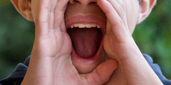
In order to breathe in, we must flatten the dome-shaped diaphragm; to breath out, we let it relax again. The diaphragm delivers oxygen to us a dozen times or more each minute, a half-billion times during an 80-year life.

"We are completely dependent on the diaphragm," said Gabrielle Kardon, a biologist at the University of Utah. "But we take it for granted every moment we're breathing."
To Dr. Kardon, the diaphragm is not just underappreciated but puzzling. All mammals, from platypuses to elephants, have a diaphragm. But no other animal has one. "We have a very different solution for breathing than reptiles and birds," said Dr. Kardon.
Before the evolution of a diaphragm, our reptilelike ancestors probably breathed the way many reptiles do today. They used a jacket of muscles to squeeze the rib cage.
Once the diaphragm evolved, breathing changed drastically. Mammals gained a more powerful, efficient means to draw in a steady supply of oxygen. The evolution of a diaphragm may thus have made it possible for mammals to then evolve a warm-blooded metabolism. Without a diaphragm, humans might not have been able to evolve giant - but oxygen-hungry - brains.
Scientists suspect that the diaphragm evolved through some change in the way mammal embryos develop: Mutations caused certain embryonic cells to grow into an entirely new muscle. Dr. Kardon and other researchers are trying to understand that shift and why the muscle sometimes fails to develop, with catastrophic consequences.
One in every 2,500 babies is born with a hole in its diaphragm. The baby's liver, intestines and other abdominal organs can push up through this opening against the lungs, stunting their growth and restricting the baby's breathing. About a third of babies born with congenital diaphragmatic hernias die, and it is likely that still more die of this defect before birth.
Scientists have found that mutations in certain genes can increase the risk of developing hernias. But they have struggled to figure out exactly how these genes build the diaphragm. Dr. Kardon and her colleagues recently developed a set of new tools get a closer look. They published the research March 25 in Nature Genetics.
They engineered mice so that certain types of cells would glow inside mouse embryos. Then they tracked the cells as they multiplied and migrated.
The diaphragm begins as a pair of folds flanking the esophagus, she and her colleagues found. These folds then expand in two waves. "It's beautiful, aesthetically," said Dr. Kardon.
In the first wave, one set of cells in the folds multiplies outward, toward the sides of the body. Then these cells fan out toward the front and back. The cells become connective tissue, forming a thin membrane across the top of the liver.
In the second wave, muscle-generating cells emerge from the folds. They follow the trail blazed by the connective tissue, forming a second sheet sandwiched inside the membrane. "The muscle cells are kind of dumb, and they're just following the connective tissue," said Dr. Kardon.
As part of their experiment, Dr. Kardon and her colleagues examined GATA4, a gene linked to diaphragmatic hernias. They engineered mouse embryos in which they could shut down GATA4 only in certain types of cells, and only at certain points in development.
In one trial, the scientists turned off GATA4 in the muscle cells in the diaphragm. In these cases, the mice formed diaphragms. But when the researchers shut down GATA4 in the connective tissue, the mice developed hernias.
Connective tissue cells must be using GATA4 to lay down a chemical trail for muscle cells, Dr. Kardon concluded. They can still lay down the trail if they have one defective copy of the GATA4 gene.
Each time the connective tissue cells divide, there is a chance that a working copy of GATA4 may mutate, too. If that happens, the mutant cell and its descendants can't lay down a trail, resulting in a gap in the sheet of muscle.
As the liver pushes against the diaphragm, the pressure creates intense stress in the gap, causing the diaphragm to rupture.
Clifford J. Tabin, a geneticist at Harvard Medical School, said that the new study offers a molecular explanation for how congenital diaphragmatic hernias occur. "I think it is a beautiful study and terribly important," he said.
John J. Greer, a biologist at the University of Alberta, said he was skeptical that this scenario could account for most hernias.
He noted that most medical cases of congenital diaphragmatic hernias occurred in the back left or right corners of the diaphragm. Dr. Kardon and her colleagues produced many hernias in the middle or front of the diaphragm of their mouse subjects.
Dr. Kardon countered that a lot of hernias occur in other parts of the diaphragm, but doctors fail to notice many of them. because the lungs sit at the back of the diaphragm, hernias there can be dangerous. Hernias elsewhere can be harmless.
"Because they don't have serious medical consequences, they go unnoticed," she said.
It is possible that the diaphragm may have evolved in two waves, much as it develops in the embryo. Dr. Kardon suggested that the ancestors of mammals may have evolved a connective tissue sheet first, simply to separate the lungs from the abdomen.
Only later did muscles form a sandwiched layer, creating a breathing pump.
This transition may not have required a lot of mutations. Muscle cells follow chemical trails made by connective tissue in other parts of the body. Once the connective tissue cells started making a proto-diaphragm, muscle cells already had the molecular machinery required to follow them.
"The mechanism is already in place," said Dr. Kardon. "It's a relatively simple step, even if it sounds like an impossible chasm."
(Source: New York Times)