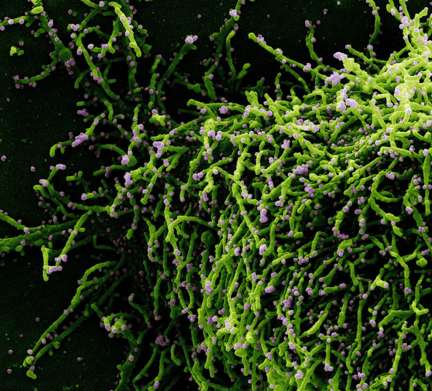
Researchers have now published a study that has revealed how SARS-CoV-2, the virus that causes COVID-19, can directly infect brain cells called astrocytes. This work has also demonstrated that even mild cases of COVID-19 may cause structural changes in the cerebral cortex, the thin outer layer of the brain that has a central role in many functions of the human brain. This research has shown that SARS-CoV-2 uses a protein called neuropillin to invade astrocytes, which is different from the ACE2 receptor the virus is known to use to infect cells in the nose and lungs. The findings have now been reported in the Proceedings of the National Academy of Sciences.
There has been ongoing debate about how SARS-CoV-2 impacted the brain, and whether the virus itself was directly infecting brain cells, or if side effects of the infection such as blood vessel damage were affecting the brain.
While two other studies have shown that SARS-CoV-2 viral particles could be found in the brains of some COVID-19 patients, people were still unsure about whether it was in the blood supply, endothelial cells lining blood vessels, or nerve cells. This study has shown for the first time that the virus does infect astrocytes in the brain directly and can replicate inside of them, which reduces the viability of neurons, explained study co-author Daniel Martins-de-Souza, a professor at UNICAMP's Biology Institute.
Astrocytes are commonly found in the central nervous system, and while they were once thought of primarily as support structures for neurons, researchers have found that they have other crucial roles; they can help regulate the levels of important molecules such as neurotransmitters, and assist in maintaining the blood-brain barrier.
In this study, the researchers analyzed brain tissue from 26 people who had died of COVID-19. The virus was detected in astrocytes in the brains of five of the individuals. There were also structural changes that may have indicated damage to the central nervous system.
"We observed signs of necrosis and inflammation, such as edema, neuronal lesions, and inflammatory cell infiltrates," said study co-author Thiago Cunha, a professor at FMRP-USP.
The research team also confirmed that SARS-CoV-2 can infect astrocytes, by collecting human brain tissue during brain surgeries, and exposing it to the virus in the lab. There were also biochemical changes in the infected astrocytes that are associated with metabolism. Studies of astrocytes in culture suggested that the mitochondria in these infected cells were not functioning correctly. The infected astrocytes were using more glucose than usual, but their energy output was not increasing.
The mitochondrial dysfunction in SARS-CoV-2-infected astrocytes could be altering the levels of certain neurotransmitters including GABA. GABA is knowno to normally promote relaxation and calm by preventing excessive firing in neurons.
The researchers observed increased cell death when neurons were grown in culture with media from infected astrocytes.
The investigators also examined 81 people with mild cases of COVID-19 using magnetic resonance imaging (MRI). COVID-19 cases were confirmed with RT-PCR, and none of the participants had been hospitalized for the illness. People who had experienced a case of COVID-19 volunteered for the MRIs an average of 60 days after the start of their infection, with about one-third of study participants still experiencing some neuropsychiatric or neurological symptoms, such as headache, fatigue, anxiety, loss of smell or taste, memory changes, and daytime drowsiness.
Data from the MRIs was compared to MRI data collected from healthy individuals. This research showed that there was a decrease in cortical thickness in some of the people with mild COVID-19 compared to the average thickness of uninfected, healthy people. The areas with diminished thickness have also been linked to some of the symptoms that people were feeling.
"We observed atrophy in areas associated, for example with anxiety, one of the most frequent symptoms in the study group," said study co-author Clarissa Yasuda, a professor and member of the Brazilian Research Institute for Neuroscience and Neurotechnology (BRAINN). "Considering that the prevalence of anxiety disorders in the Brazilian population is 9 percent, the 28 percent we found is an alarmingly high number. We didn't expect these results in patients who had had the mild form of the disease."
People with mild COVID-19 also did not do as well as the national average on tests that measure cognitive function.
Astrocytes do not carry ACE2 receptors. This study showed that SARS-CoV-2 can bind to a protein called neuropilin, found on astrocyte membranes, to infect the cells. But the researchers are still determining how the virus gets to the brain. "Some animal experiments suggest the virus can cross the blood-brain barrier. There's also a suspicion that it infects the olfactory nerve and from there invades the central nervous system. But these are hypotheses for now," said Martins-de-Souza.
What remains to be seen is how long symptoms will last for people that continue to experience what is now known as long COVID.
Experienced research scientist and technical expert with authorships on over 30 peer-reviewed publications, traveler to over 70 countries, published photographer and internationally-exhibited painter, volunteer trained in disaster-response, CPR and DV counseling.