Tuberculosis is one of the top ten causes of death worldwide, with 10.4 million people falling ill and 1.7 million dying from the disease in 2016. OF these deaths 95% of tuberculosis-related deaths occurred in low and middle-income countries with 64% of total cases arising in India, Indonesia, China, Philippines, Pakistan, Nigeria, and South Africa. A significant cause for concern is multidrug-resistant tuberculosis (MDR-TB), which remains a global public health crisis and security threat with an estimated 600,000 new cases in which resistance to the first-line drug rifampicin occurred. Currently, the Sustainable Developments Goal includes ending the tuberculosis epidemic by 2030, a lofty task despite the 2% per year decline in cases.
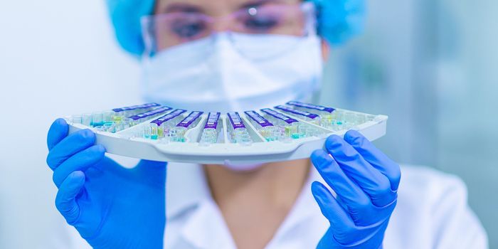
Tuberculosis is an infectious disease caused by the bacterium Mycobacterium tuberculosis that affects the lungs. These bacteria can be spread via coughing or sneezing when tiny droplets are released into the air and inhaled by another individual. Individuals may harbor the bacteria but show no symptoms, latent tuberculosis, or can present symptoms in active tuberculosis. Symptoms of tuberculosis include chest pain, persistent coughing and coughing up blood, fever, chills, loss of appetite, and many others. While a healthy immune system can successfully fight the bacteria, risk factors that weaken your immune system such as HIV/AIDS, cancer, diabetes, and malnutrition can increase the risk for the disease. Current treatment of tuberculosis includes long-term antibiotic courses of six to nine months with drugs such as rifampin, isoniazid, and pyrazinamide. If an individual has drug-resistant tuberculosis, a combination of antibiotics may be necessary.
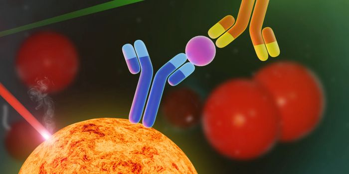
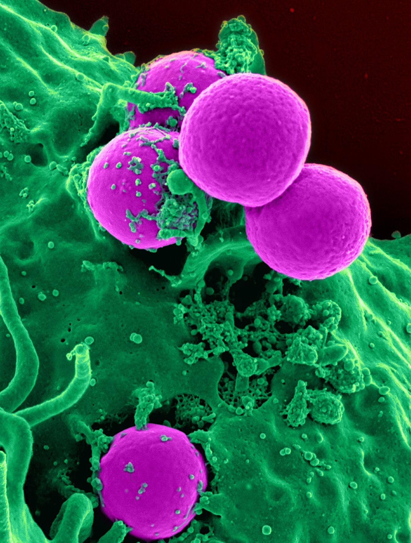
A recent study in PLOS Pathogens, by researchers at the Rutgers New Jersey Medical School, examined the underlying mechanism behind harmful white blood cell formation during tuberculosis infection. A particular type of white blood cell called a foam cell, also known as macrophages, accumulate lipids. When these cells accumulate, they contribute to a maladaptive immune response that can lead to chronic inflammation and tissue damage in various disease such as tuberculosis.
The researchers observed the formation of the foam’s cells in lung tissues of rabbits, non-human primates, and humans with tuberculosis. The study found that triglycerides, a type of fat, accumulates to form foam cells during tuberculosis infection. The formation of the triglyceride accumulation is mediated by distinct signaling pathways that are different than other disease mechanisms. "The finding that foam cells found in tuberculosis differ from those present in atherosclerosis," states the first author of the study, Valentina Gennaro, "demonstrates that these dysfunctional cells, which are associated with many diseases, are generated through biological processes that differ from one disease to another."
The identification of the molecules and pathways involved in triglyceride synthesis could provide novel biomarkers for progression of active tuberculosis. Previous research has been done on foam cells in diseases such as cancer, heart disease, and metabolic disease. Pharmacological compounds previously used as therapies against other diseases involving foam cells could be repurposed for treatments against tuberculosis.
To learn more about Tuberculosis watch the video below!
Sources: PLOS Pathogens, World Health Organization, Mayo Clinic, American Lung Association