While most traditional imaging techniques fall short for scientists looking to predict the effects of cancer immunotherapy treatments, a new method provides a unique opportunity to anticipate a person’s response and alter their medication accordingly.
Immunotherapy doesn’t work for everyone, and the risk of serious side effects makes the “just try it and see what happens” approach seem harsher than it needs to be. A doctor doesn’t want the patient to endure side effects if the treatment isn’t doing anything to kill cancer cells. With a new method capable of recording progress early during treatment, doctors can know when to push through and when to give up and try something else.
A new noninvasive PET imaging method is the newest beam of hope, and the subject of serious research interest at the American Association for Cancer Research. Unlike traditional techniques like CT, MRI, and FDG PET scans which can’t tell the difference between a tumor growing despite immunotherapy treatment and a tumor growing because of immunotherapy treatment, noninvasive PET imaging provides a clear distinction. While immunotherapy doesn’t cause tumor cells to proliferate - that would be counterintuitive - the treatment does bring in large populations of immune cells and increase glucose uptake, making the tumor appear on traditional images as just as large as a tumor unaffected by the treatment.
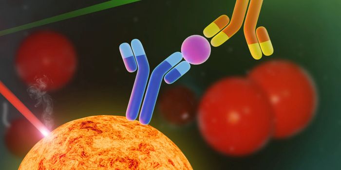
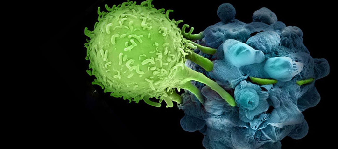
The method works by measuring granzyme B, a serine protease released by immune cells to target and kill tumor cells. Using a probe that binds granzyme B once it is released, PET scanning noninvasively provides images of where in the body immune cells are unleashing granzyme B on cancer cells.
A research team from the American Association for Cancer Research tested the probe and PET method in mice with tumors treated with an immunotherapy based on checkpoint inhibitor drugs.
"Because PET imaging is quantitative, we could measure the degree of effectiveness and put a number on it," explained Umar Mahmood, MD, PhD. The mice whose immune cells released large amounts of granzyme-B as detected by PET imaging were the same ones who responded to immunotherapy over time and showed a reduction in tumor size. The mice whose immune cells produced little granzyme-B were not as lucky.
Next, Mahmood also tested the probe technology with nine human melanoma biopsy samples and saw similar results.
“The ability to differentiate early in the course of treatment patients who are likely to benefit from immunotherapy from those who will not can greatly improve individual patient care and help accelerate the development of new therapies," Mahmood said.
The present study was published in the journal Cancer Research.
Sources: American Association for Cancer Research