The immune response needs to be just right: not too much, or an autoimmune disease will develop; not too little, or viruses and bacteria will swarm the body and wreak havoc. The body has regulatory mechanisms in place to maintain this delicate balance, and University of Alabama at Birmingham (UAB) scientists, in a new Nature Immunology study, recently examined the intricate details.
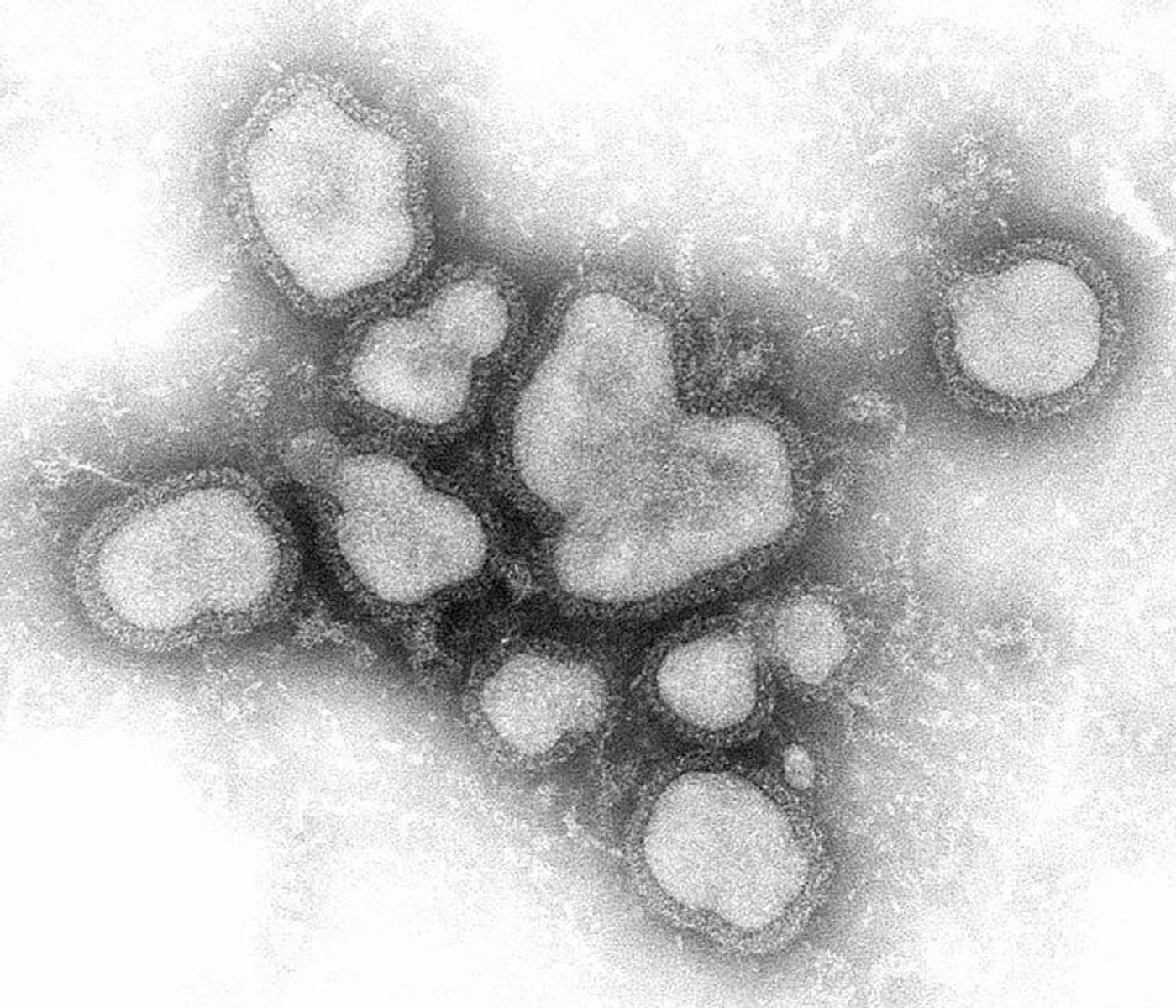
Using a mice model of influenza infection, researchers mapped out the regulatory mechanisms employed by the immune system to prevent autoimmune reactions leading to diseases like multiple sclerosis, lupus, and rheumatoid arthritis.
First, they saw that T follicular regulatory cells (TFRs) stand out against other regulatory T cells, called Tregs; they develop during the later stages of the immune response to influenza infection as opposed to during the beginning of the infection.
However, there’s a purpose for this delayed development.
As the immune response to influenza begins, levels of a signaling molecule called interleukin-2 (IL2) start to increase, which triggers the production of Tregs. These cells are charged with the duty of making sure the immune response doesn’t go overboard; once the virus is cleared out, the immune system eases up.
At the same time, IL2 signaling prevents TFR cells from developing during the peak of the immune response. But by the end of the battle versus the influenza virus, Tregs suppress activity of IL2 signaling and transform into TFR cells.
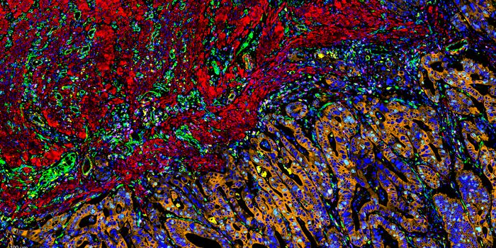
TFR cells then travel to the follicles of the lymph nodes, also the place where B cells produce antibodies which replicate and, sometimes, mutate. However, TFR cells seemed to serve the purpose of preventing the collection of B cell variants that were producing autoreactive antibodies. TFR cells left B cells producing influenza-targeting antibodies alone.
When researchers removed TFR cells from the situation, there was nothing standing in the way of misguided B cells creating antibodies that target the body’s own cells.
TFR cells were first identified in 2011 as a unique subset of Tregs, and they’ve been found to suppress T follicular helper cells in addition to B cells. T follicular helper cells join B cells in promoting antibody production. UAB’s André Ballesteros-Tato, PhD, recognizes the role of TFR cells as an important part of understanding how the immune system is regulated: "this research gives us clues of what to look for when we look at how autoimmune disease develops.”
Sources: Trends in Immunology (1, 2), University of Alabama at Birmingham