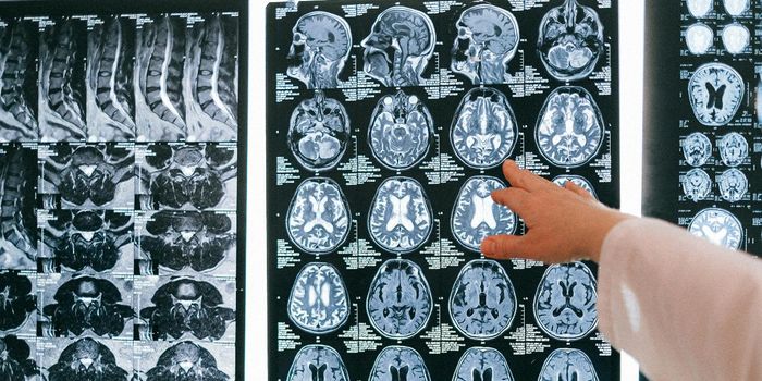
While doctors, scientists and medical researchers have good imaging tools to see blood, bones and muscle, seeing what happens in the brain is not as straightforward. While tests like CT scans, PET scans and MRIs are helpful, they can’t show what is happening in the brain at the cellular level. A new study and device being developed in Boston might be just what is needed to get a good look at brain activity.
A study detailing the findings of a group of neuroscientists at Harvard University was published recently in the Journal Cell and the findings were unprecedented. Jeff Lichtman,
the senior study author said in a press release “I'm a strong believer in bottom up-science, which is a way of saying that I would prefer to generate a hypothesis from the data and test it. For people who are imagers, being able to see all of these details is wonderful and we're getting an opportunity to peer into something that has remained somewhat intractable for so long. It's about time we did this, and it is what people should be doing about things we don't understand.”
In the study Lichtman and his team took ultra-thin slices of mouse brains and were able to create images of the brain structures at a nanoscale resolution. The team started with the area of the mouse brain that receives input from the whiskers of the mouse. Mice use their whiskers to make sense of the space around them, to find their way in and out of small areas and the sensitivity of these whiskers is very high.
To create images of different parts of the sensory input such as neurons, glial cells, blood vessels and the like, study co-author Daniel Berger of Harvard and MIT created a program called VAST that assigned colors to each brain piece to set them apart from each other so they could be seen individually.
While the images were complex and unlike anything seen before, the real significance of the study is that the team took these images and from them was able to construct an incredibly detailed picture of what was happening in a very tiny space.
The sample size was only about 1500 cubic microns, however using electron microscopy and the VAST program the team was able to identify over 1700 different synaptic connections and image all the parts and pieces that went into each. The amount of detail from such a small sample, represented in such high resolution has never been accomplished before this study.
Another finding from this research was in the area of axonal transport. While so much is going on, it’s not random. Rather than axons transporting synaptic signals along to dendrites near them, there were specific patterns that emerged. Location was not the determining factor in which dendrites would connect with which axon.
Narayanan "Bobby" Kasthuri, of the Boston University School of Medicine who was the study’s first author
talked about the patterns found and said, "We had this clean idea of how there's a really nice order to how neurons connect with each other, but if you actually look at the material it's not like that. The connections are so messy that it's hard to imagine a plan to it, but we checked and there's clearly a pattern that cannot be explained by randomness."
Check out the video below to see the team explain some of their research and what they think it means for the field of neuroscience.
_ffe232d2eb9981d7235e41e4e6b2a1fc.jpg)