While there are many ways to image the brain including CT scans, PET scans and MRIs, the best way to observe and monitor brain activity is by directly placing electrodes and nanowires into brain tissue. Of course this has consequences such as tissue damage, infection and even bleeding. While some electrodes are very small, and wires can be extremely thin, brain monitoring apparatus is difficult to use and balancing the benefit against the risks has been a challenge for neuroscientists.
Researchers at Lund University in Sweden may have a solution. A team of scientists at the Department of Experimental Medical Science, at the Neuronano Centre at the university have been working for almost nine years on electrodes that can be implanted in the brain on a long-term basis. It’s no easy task, but a paper recently published in the journal Frontiers of Neuroscience details their project which includes the development of electrodes that can capture signals from single neurons in the brain over time and yet still not cause brain tissue damage.
Professor Jens Schouenborg led the project together with Dr Lina Pettersson. If their prototypes work and can be tolerated by human patients it would go a long way towards understanding brain function not only in patients with neurological conditions, but in healthy subjects as well.
In a press release from the Lund University Professor Jens Schouenborg said, “There are several elements that must go hand in hand for us to be able to record neuronal signals from the brain with decisive results. First, the electrode must be bio-friendly, that is, we have to be confident that it does not cause any significant damage to the brain tissue. Second, the electrode must be flexible in relation to the brain tissue. Remember that the brain floats in fluid inside the skull and moves around when we, for instance, breathe or turn our heads. The electrode and the implantation technology that we have now developed have these properties, which is unique.”
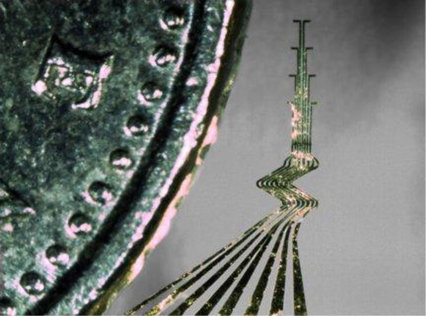
The electrodes, which the Lund team calls “3-D Electrodes” are unlike any others. They are made so that they can be flexed in all three dimensions, and are extremely soft. This kind of construction makes it possible for the electrodes to stay in the brain tissue longer than other stiffer models. It’s a matter of getting the technology just right. Electrodes that are too flexible will not hold their position in the brain. Those that are too stiff can cause tissue damage, cell death and other complications.
They start out as very thin slices of gold measuring just 10 microns in thickness. Using a laser cutter, they cut into very precise specific shapes and insulated with a polymer called parylene C. Finally, they are coated in layers of gelatin to make them firm enough to be inserted into the brain. Once there, the gelatin coating dissolves and the electrode is left in place.
The Lund team hopes to build on their research which used lab rats as subjects and develop the electrodes for use in human trials. Check out the video below to see more about this advance in neuroscience.