The vast network of neurons, dendrites, axons and other hardware that is responsible for sending messages all over the body is incredibly complex. Nothing can work without this supercomputer processing and sending signals in exactly the right order and time. It’s an area of neuroscience that researchers are constantly studying. If more can be found out about how it works, then treatments can be found for when it doesn’t work properly.
One of the recent advances in this area comes from Columbia University scientists who have developed a new optical technique to study how information is transmitted in the brains of mice. With so many signals zipping through the brain, they expected that there were be many connections throughout the brain. These connections are called synapses. However, what they found with this method was not what they expected. Only a small portion of synapses are active after the mouse brains were electrically stimulated.
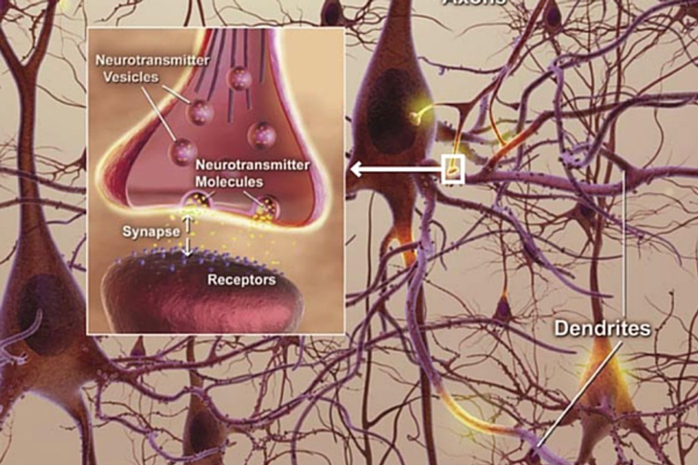
In a press release from Columbia, one of the study authors, David Sulzer, PhD, professor of neurobiology in Psychiatry, Neurology, and Pharmacology at Columbia University Medical Center (CUMC) said, “Understanding how we accomplish complex tasks, such as learning and memory, requires us to look at how our brains transmit key signals—called neurotransmitters—across synapses from one neuron to another. Older techniques only revealed what was going on in large groups of synapses. We needed a way to observe the neurotransmitter activity of individual synapses, to help us better understand their intricate behavior.”
But how could they get a view at this tiny level? The team worked with another lab at Columbia, run by of Dalibor Sames, PhD, associate professor of chemistry. They developed a novel compound called fluorescent false neurotransmitter 200 (FFN200). It gets added to brain tissue or nerve cells in mice and when it interacts with the synaptic activity it fluoresces and gives a live action picture of brain messaging as it happens.
Dopamine, the key neurotransmitter involved in learning, habits and reward seeking, was viewed as it was released and taken back up using a fluorescence microscope. They expected to see that all the synapses that were electrically stimulated would light up and release dopamine. Only about 20% of them actually did.
Researchers hope to do more work on why so many of the synapses didn’t activate. Neurological diseases like Parkinson’s, Alzheimer’s and schizophrenia all involve dopamine disruption to a certain extent so understanding how these patterns function could lead to treatments for those conditions.
The research is published in the journal
Nature Neuroscience. Check out the video below to see how the imaging worked and hear more about the research.