Picture a lab, with a semi-evil looking scientist cackling about controlling the brain with pulses of light. Sound like the newest science fiction blockbuster? No, it’s the exciting field of optogenetics. In neuroscience research, seeing how the brain works is crucial and extremely difficult.

It’s not like the heart or kidneys where MRI scans and blood tests can show the structure and function of those vital organs. While MRI technology as well as PET scans, CT scans, molecular imaging, photon and electron microscopy and so much else are valuable tools, they can only go so far.
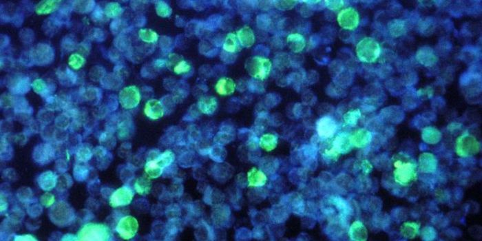
Optogenetics to the rescue? Well, in the last decade or so, it’s been refined and used in many studies. Most scientists in the field agree that the first major work to talk about optogenetics and demonstrate what it could do was the study published in 2005 by Stanford scientist Karl Deisseroth and MIT professor Ed Boyden. Another neuroscientist, Dr. Xhou-Hua Pan at Wayne State University was conducting similar work, which was later published as well. Their work, with the protein channelrhodopsin-2, a photoreceptor, showed how neurons in the brain could be manipulated by light. Passing light through a particular cell can change the electrical charge of that cell and cause it to fire on command. Getting individual brain cells, called neurons, to fire in specific ways was a huge breakthrough in neurological research.
The brain is essentially a collection of tiny but extremely active electrical circuits. Charged ions fly around these circuits, causing neurons to fire and messages from one part of the brain to be transmitted to another part of the brain. It’s this transmission that is key to everything the brain does. When injury or illness is a factor, being able to manipulate this activity could be the way to treat patients and give them back what the injury or illness has taken away.
Channelrhodopsin-2 was found to be a key player in opening ion channels in the neural networks of the brain. Since it’s light reactive, it can switch cells on and off. Getting it into the cell was a bit complex. Using viruses, channelrhodopsin-2 was inserted into individual neurons. When light was passed through the neurons that had channelrhodopsin-2 inserted, they fired, on command, and optogenetics was born.
Since then, genetic engineering has allowed for the creation of mice that will naturally express channelrhodopsin-2 in their neurons. These are the lab animals used in many studies that involve investigation of optogenetic technologies. Tiny fiber optic wires are then inserted into the brains of these mice to stimulate whatever region they want to look at. These manipulations have varied from attempting to turn off a gene thought to cause dementia to inhibiting or sparking certain behaviors. Channelrhodopsin-2 is the only gene necessary for optogenetics to work, so it allows researchers to look at any of the thousands of neurons in the brain.
The video explains the role of optogenetics in neuroscience. It’s a technology that’s likely to be at the forefront of neuroscience research for years to come, so check it out.
Sources: Scientific American, The Scientist.com, The New Yorker, The Max Planck Institute