
Director of Applications, Veranome Bio
BIOGRAPHY
Speaker
Abstract
Single-cell spatial omics is a burgeoning area of research that has been generating incredible excitement in academic, translational and clinical circles. One of the biggest challenges in this area is producing and subsequently integrating hundreds of overlapping images that contain thousands of nanoscale signals against the noisy background of a cell. The Pisces workflow robust, easy-to-use, end-to-end multi-omics solution for highly multiplexed targeted Spatial RNA analysis. VeranomeBio’s Pisces workflow consists of 1) a novel assay for the massively parallel imaging >100s of RNA targets, 2) state-of-the-art imaging technology delivering sub-cellular resolution, and 3) a software suite for from custom panel design, experimental setup and data processing, as well as sophisticated visualization and statistical analytics of segmented cell results. The presentation includes detailed analysis of human and mouse tissues, including tumor samples, mouse neural tissues and organoids. We enable researchers to deepen their understanding of diseases with high-quality data of how individual cells organize within tissue, using robust workflows, reliable imaging systems and without the need for extensive bioinformatics tools or infrastructure. In this study, we demonstrate the results with mouse liver and neural tissues, as well as the spatial organization of differing cell layers composing the structure of a newly harvested organoid. Initiating with a PFA-fixed tissue mounted in a proprietary flow cell, a targeted panel is used to localized individual RNA molecule within specific compartments within a cell. Through cyclic reagent addition and imaging, the barcoded panel enables the Pisces system to reveal and decode custom or pre-validated panels tied to specific disease states or biological functions. Individual cells are identified through advanced, AI-based segmentation algorithms, and individual RNA molecules are quantified within cell boundaries. The resulting matrix of individual cells, summarized RNA counts, coupled with X, Y and Z coordinates are used for statistical clustering and phenotyping, as well as spatial relationships characterizing the molecular and cellular niches and architecture. The Veranome system provides the first commercial system for high-content imaging and analysis of true single cell, spatial transcriptomics.
Learning objectives:
1. Demonstrate the latest solution for a high-plex spatial transcriptome analysis of complex tissues at single cell resolution
2. Image processing through data visualization of complex data sets for in situ characterization of organoids and tumor/normal tissues
3. Critical components for sub-cellular analysis including sample preparation, imaging hardware, image analysis and cell phenotyping methods
You May Also Like
JAN 14, 2026 | 10:00 AM

Join us for an insightful webinar to discuss a holistic approach to testing for TB in a clinical setting. Tuberculosis remains a global health challenge and accurate, timely diagnosis is cri...
JAN 15, 2026 | 8:00 AM
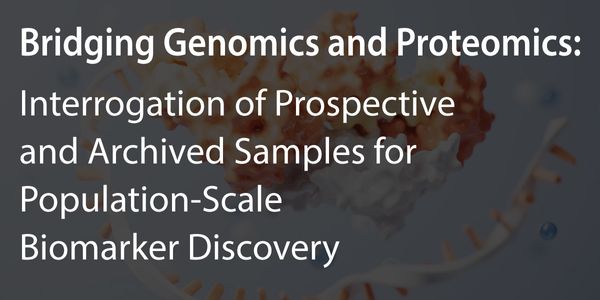
Personalized medicine promises to significantly improve patient outcomes, but achieving this requires a deep understanding of human health and disease mechanisms at the molecular level. The...
JAN 20, 2026 | 11:00 AM

CAR-T translational research demands precise, reproducible, and scalable flow cytometry workflows. Manual centrifugation and antibody preparation steps remain major sources of variability an...
JAN 20, 2026 | 9:15 AM
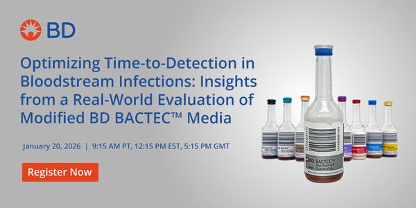
Bloodstream infections (BSIs) remain a critical challenge in clinical care, where every hour of delay in diagnosis can significantly impact patient outcomes. This webinar explores findings f...
JAN 21, 2026 | 8:45 AM
C.E. CREDITS

Lipid nanoparticles (LNPs) have become the gold standard in non-viral gene delivery technologies, exemplified by the approval of the LNP messenger RNA (mRNA) vaccines against SARS-CoV-2. Sin...
JAN 22, 2026 | 8:00 AM
Cell line development (CLD) is often a critical bottleneck in biopharmaceutical production, often requiring labor-intensive workflows and multiple sequential screening steps that extend time...
Loading Comments...