While cholesterol is primarily found in the cell membrane, its role there is not well understood. Researchers at the University of Illinois at Chicago have used a powerful imaging technology to reveal more about the function of cholesterol. In a surprise to the investigators, cholesterol apparently acts to transmit signals across the membrane of cells. Their research has been reported in Nature Chemical Biology.
"Cholesterol is a lipid that gets bad press because of its association with cardiovascular disease," said the leader of the study, Wonhwa Cho, a Professor of Chemistry at UIC. "It's been very well studied, but not much is known about its cellular function. What is its role? Is it a bad lipid? Absolutely not - for example, the brain is about half lipid, and cholesterol is the richest lipid in the brain," he said.
Previous work by Cho's team has investigated the various molecular interactions cholesterol has with a variety of molecules that govern cellular processes, but researchers had not considered a regulatory role for cholesterol itself. "We knew it could play an important role in cell regulation - for example, in proliferation or development. We know that high-fat diets, which boost cholesterol levels, have been linked to an elevated incidence of cancer. How, is not fully understood," Cho explained.
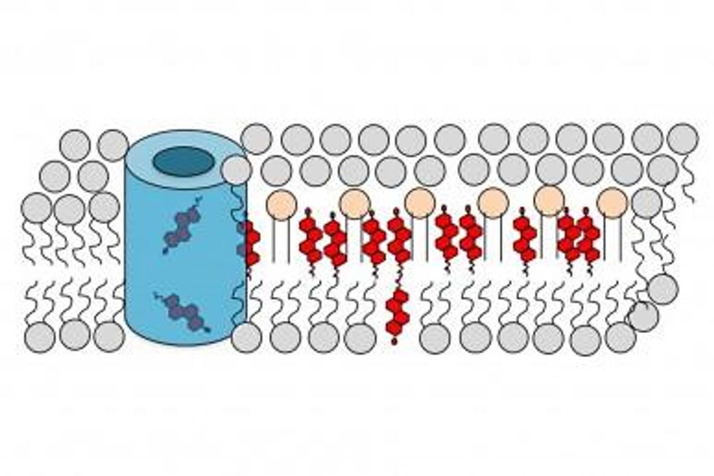
One reason that cholesterol is not considered regulatory is because such molecules are typically transitory signals. "But cholesterol is there all the time," Cho said. The majority of cholesterol in a cell, 90 percent, is found in the cell membrane, where cholesterol is a major component of membrane lipids. The cholesterol adds stability to the double-layered lipid membrane. Cholesterol coalesces as "rafts," and had been thought of as platforms used by signaling molecules. "But in this paper, we showed that a single cholesterol molecule can itself be the signal trigger," Cho explained.
It was thought that cholesterol was present in both layers of the membrane, Cho said, "maybe more in the inner layer. But we, for the first time, measured cholesterol levels in the inner and outer layers simultaneously in real time, in living cells. And we showed that cholesterol is predominantly in the outer layer."
In this work, the investigators report that cholesterol comprises around 40 percent of the membrane’s outer layer, while the inner layer only contains around 3 percent cholesterol. The various levels of cholesterol can be altered; the amount in the inner layer more than doubled and the level in the outer layer fell by that amount after a stimulation by the researchers.
They also determined that in cancerous cell, the normally low level of cholesterol in the inner layer jumps much higher. "We checked this in many different cell lines," Cho added.
Statin drugs, used to lower cholesterol, have the side effect of reducing the risk of cancer. Cho’s team determined that applying a statin to cells substantially lowered the amount of cholesterol in the inner layer, reducing cellular growth. This could potentially lead to new cancer treatment methods, Cho suggested.
"I think we're just scratching the surface of the regulatory role of cholesterol. We have many unpublished data indicating that cholesterol is involved in a wide variety of cellular processes and regulation," he said.
Cho added that cholesterol is a challenging molecule to study; lipids like cholesterol are "very nasty molecules to work with; unlike most biological chemicals, they are not soluble in water. "We had to devise a new strategy," Cho explained.
Cho and his team created an optical imaging technology six years ago, which enables the direct measurement of lipid levels in live cells. By adding a fluorescent tag to a lipid-binding protein molecule, the amount of free to bound lipid can be measured, as there is a change of color when the lipid is in specific locations within the membrane.
To learn more about the fluid mosaic model of the cell membrane, check out the video above from Khan Academy. The video below is a short lecture on glycolipids and cholesterol.
Sources: AAAS/Eurekalert! via UIC, Nature Chemical Biology