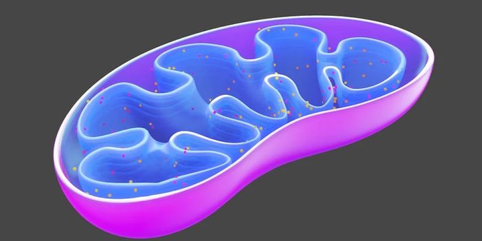
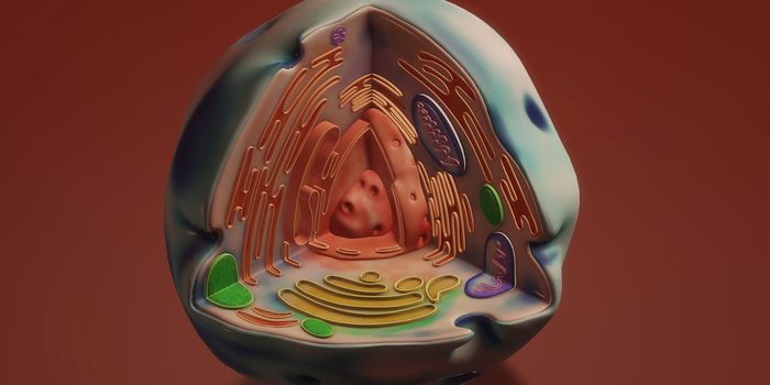
Our bodies replace millions of cells every day; to do so, a cell has to divide into two new daughter cells. The process requires replicating the entire genome and dividing the DNA correctly between the two new cells. Our genome can get damaged as this happens, but it can also acquire damage from many different environmental influences. Scientists have now discovered how two proteins can stabilize DNA that’s been damaged, and protect the integrity of the genome in the process. Their work, which has been reported in Nature, also shows how people that carry defects in some proteins are more susceptible to disorders like cancer, dementia, or immune problems because they can’t maintain stability in their genome or repair damaged portions.
Two proteins, 53BP1 and RIF1, were found to generate a three-dimensional scaffold around DNA strands that have broken. This scaffolding concentrates scarce repair proteins, and the critical work of DNA repair can then begin. This ensures that DNA stays stable, and prevents more damage from occurring in the surrounding area.
“It's a unique discovery. Understanding the body's natural defense mechanisms enables us to better understand how certain proteins communicate and network to repair damaged DNA. This opens up an opportunity to better design how DNA damage causes disease and design drugs that improve treatment of patients with unstable DNA,” said study co-author Professor Jiri Lukas, Center Director at the Novo Nordisk Foundation Center for Protein Research.
In this study, the researchers applied cutting-edge super-resolution microscopy that enabled them to visualize the protein scaffold as it assembled around the DNA break.
“This could be compared to putting a plaster cast on a broken leg; it stabilizes the fracture and prevents the damage from getting worse and reaching a point where it can no longer heal,” said the first author of the study, postdoctoral fellow Fena Ochs, of the Novo Nordisk Foundation Center for Protein Research.
It had been thought that these proteins were only acting in the area where DNA strands were broken. Their investigation allowed the researchers to see the large structure that 53BP1 and RIF1 formed around the breaks.
“Roughly speaking, the difference between the proportions of the protein-scaffolding and the DNA fracture corresponds to a basketball and a pinhead,” Ochs noted.
The researchers suggested that the protein scaffold shows that the DNA break must be stabilized, and so does the area around it. The damaged area is this preserved, there is less of a chance that more damage will occur, and the special proteins doing the repair, members of the shieldin network, can do their jobs.
When the scaffolding proteins were removed, the researchers saw that large sections of the chromosome around the break rapidly fell apart. When the scaffolding proteins are absent, cancer can arise, because although the cells attempt to fix the problem, the effort is futile. People that don’t carry functional copies of these proteins are more likely to get diseases that arise from unstable DNA.
Sources: AAAS/Eurekalert! via University of Copenhagen, Nature