
Professor, University of British Columbia
BIOGRAPHY
SEP 17, 2025 9:00 AM PDT
Keynote Presentation: SuperResNET: Learning in-cell macromolecular architecture from SMLM data w/ Live Q&A
Presented at:
Cell Biology Virtual Event Series 2025
Speaker
Abstract
Protein structures are now being resolved at the atomic level, but deciphering their molecular organization in the cell remains a challenge. Super-resolution microscopy enables the use of fluorescent-based molecular localization tools to study molecular structure at the nanoscale level in the intact cell, bridging the mesoscale gap to classical structural biology methodologies. SuperResNET is an integrated machine learning-based analysis software for visualizing and quantifying 3D point cloud data acquired by single molecule localization microscopy (SMLM). The computational modules of SuperResNET include correction for multiple blinking of a single fluorophore, denoising, segmentation (clustering), and feature extraction, which are then used for cluster group identification, modularity analysis, blob retrieval and visualization in 2D and 3D. More recent updates to SuperResNET allow two-channel interaction distance analysis to determine how two proteins interact within macromolecular assemblies. SuperResNET can be effectively and easily applied to any SMLM event list from which it rapidly learns macromolecular architecture in the intact cell. I will describe the ability of network graph analysis software (SuperResNET) to determine molecular structure from dSTORM and MinFlux single molecule localization microscopy. Use cases to be described include molecular analysis of the nucleopore complex, structural changes to clathrin coated pits by inhibitors of clathrin endocytosis and the identification and structural characterization of caveolae and non-caveolar caveolin-1 scaffolds.
Learning Objectives:
1. Explain how SMLM breaks the diffraction barrier
2. Compare structure determination by SuperResNET to that obtained by cryoEM
3. Describe the structure and function of Cav1 scaffolds and caveolae
You May Also Like
FEB 17, 2026 | 8:00 AM

In cell therapy applications, commonly employed cell purification platforms for large-scale isolation of immune or stem cells separate cells based on a single cell surface antigen. Enrichmen...
FEB 18, 2026 | 8:00 AM

With advancing food allergy research and emerging therapies, new allergy testing requirements arise. In this context, Basophil Activation Testing (BAT) has emerged as an essential component...
FEB 18, 2026 | 1:00 PM

Energy-efficient filtration solutions that reduce hidden chemical exposure from benchtop work and lab equipment to improve overall laboratory safety...
FEB 19, 2026 | 7:00 AM
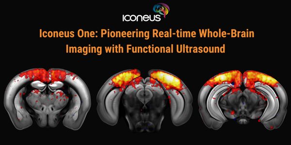
Discover how Iconeus One, the premier functional ultrasound (fUS) system for neuroscientists, is transforming brain function studies. Based on ultrafast plane-wave sonography, Iconeus One of...
FEB 19, 2026 | 8:00 AM

The collection of high-quality genomic DNA remains a major barrier in pediatric and neurodevelopmental research, particularly among children with autism spectrum disorder (ASD) and other neu...
FEB 23, 2026 | 9:00 AM
C.E. CREDITS

Explore decentralized liquid biopsy with SOPHiA GENETICS and MSK-ACCESS®, expanding access, improving outcomes, and accelerating precision oncology....
Loading Comments...