Researchers working at the Karolinska Institutet have made a breakthrough that will aid scientists studying biofilms. They have created chemicals they call molecular chameleons, which can bind to components of biofilms and emit colors at various wavelengths as they change shape. The scientists do a very nice job describing their work in the following short video.
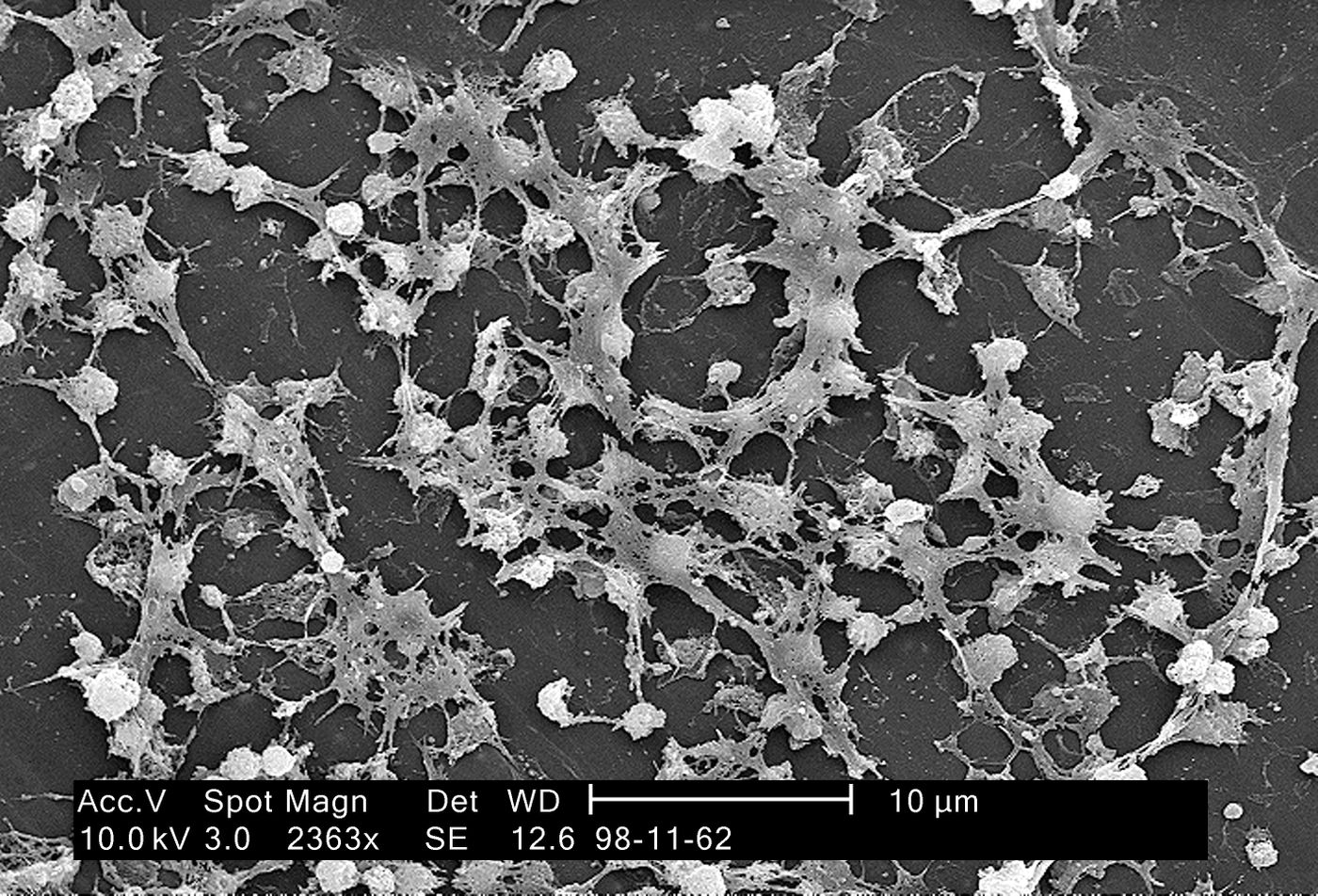
There are not a lot of good tools for researching bacterial biofilms, a colony of microbes that adheres to a surface and usually oozes a slimy matrix. Until now, there has not even been a protocol for detecting biofilms; while it is possible to see bacteria, the extracellular matrix is observed with dyes that bind in a non-specific way to molecules with charge, such as DNA and bacterial proteins. However, that inferior staining method quickly causes cell death.
Biofilms can and do form on many things, like the surface of stagnant water, medical devices implanted in patients or the tile in our bathrooms. Pathogenic biofilms can be very dangerous and are involved in many infections that cause illness. They have been implicated in health problems like gingivitis and ear infections, and must be managed during wound healing. Understandably, there is a need to understand more about them.
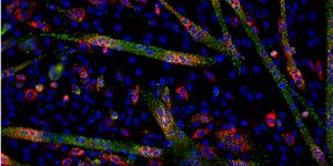
The new tool has a particular appearance depending on what they are bound to. One portion can emit light while another binds to a specific molecule in the biofilm. The molecular chameleons change color after they bind the matrix, and they are non-toxic.
"The molecules we have developed are unique in that they can send out different colors, depending on how they twist and bend. We usually call them molecular chameleons, because they change color according to the environment," explained Professor Peter Nilsson, Linköping University. His research team synthesized the tracking molecules.
"This is the first method that specifically dyes the biofilm components. This means that researchers who want to study the mechanisms behind how bacteria form biofilms now have a new tool to understand the process," said the leader of the work, Professor Agneta Richter-Dahlfors at Karolinska Institutet.
In their
report, published in Nature partner journal Biofilms and Microbiomes, the investigators show how their technique can be utilized for the study of Salmonella bacteria in both infected tissue and in the lab.
The scientists would like to see their research improve biofilm detection in biomedical research and the healthcare and food industries in order to reduce health risks to the public. There might also be applications in areas where the formation of a biofilm is a positive development, such as in the production of biogas from bacteria.
"We are very excited to be able to provide this new tool for microbial researchers to be able to use and we are looking forward to seeing new results in the near future,” concluded Professor Richter-Dahlfors.
Sources:
Emerging Infectious Diseases,
AAAS/Eurekalert! via
Karolinska Institutet,
Biofilms and Microbiomes