
Get closer to in vivo predictions with Gibco cell culture systems. Our systems allow you to closely mimic the in vivo state and generate more physiologically relevant data. Each lot of primary cells is performance tested for viability and growth potential.

-

Illumina
As a startup, Illumina aspired to transform human health. Our initial products enabled researchers to explore DNA at an entirely new scale, helping them create the first map of gene variations associated with health, disease, and drug response. Every breakthrough opened up a new ...
See more -
NanoCellect Biomedical, Inc.
NanoCellect is committed to empowering every scientist to make discoveries one cell at a time, with modern and simple technologies for cell based assays. Our microfluidic flow cytometry platforms enable biomedical scientists to analyze and sort cells required for drug discovery ...
See more -
Sartorius
The Sartorius Group is a leading international partner of biopharmaceutical research and the industry. With innovative laboratory instruments and consumables, the Group's Lab Products & Services Division concentrates on serving the needs of laboratories performing research and ...
See more -
Bio-Rad Laboratories
Bio-Rad's Single-Cell ATAC-Seq (scATAC-Seq) enables genome-wide profiling of epigenomic landscape at the single-cell level with high number of reads per cell so you can better understand the mechanisms that drive how genes are regulated.
-
MicroGem
At MicroGEM, we have re-invented nucleic acid extraction. We replace traditional extraction methods with a temperature-driven, enzymatic approach, enabling high-quality extracts from low abundance transcripts and small sample volumes. Leveraging our prepGEM Universal thermophilic ...
See more -
Cell Signaling Technology
Founded by research scientists in 1999, Cell Signaling Technology (CST) is a private, family-owned company headquartered in Danvers, Massachusetts with over 400 employees worldwide. Active in the field of applied systems biology research, particularly as it relates to cancer, CST ...
See more -
Atlas Antibodies
Atlas Antibodies is a Swedish biotechnology company driving leading research worldwide through providing affinity-purified monoclonal and polyclonal antibodies, and control antigens. Our product portfolio extensively covers human proteins in cells, tissues, and organs. With our ...
See more


SEP 26, 2019
Cell Biology Virtual Event Series 2019


The 3rd Annual Event in the Cell Biology Virtual Event Series is now available for OnDemand viewing. This years event provides an opportunity to discuss recent discoveries in biological research, advancements in techniques, and tool developments in cell research.
Cell Biology 2019 Virtual Event continues to create a valuable platform for inspiring global and interdisciplinary collaboration in a virtual environment, to study cells – their physiological properties, structure, the organelles they contain, environmental interactions, life cycle, division and death, on a microscopic and molecular level.
This years event includes the following tracks:
- Spatial Omics
- Microbiome
- Cell Biology of Genetic Diseases
Our virtual conference allows you to participate in a global setting with no travel or cost to you. The event will remain open 6 months from the date of the live event. The webinars will be available for unlimited on-demand viewing. This virtual conference also offers increased reach for the global cell biology community with a high degree of interaction through live-streaming video and chat sessions.
Continuing Education
LabRoots is approved as a provider of continuing education programs in the clinical laboratory sciences by the ASCLS P.A.C.E. ® Program. By attending this event, you can earn 1 Continuing Education credit per presentation for a maximum of 14 credits.
Use #LRcellbio to follow the conversation!
Speakers
-
Sergio Grinstein, PhD
Senior Scientist at The Hospital for Sick Children and Professor of Biochemistry, University of Toronto.BIOGRAPHY -
Zachary Bent
Director - Consumables, Product Development, 10x GenomicsBIOGRAPHY -
Sabrice Guerrier, PhD
Associate Professor of Biology, Millsaps CollegeBIOGRAPHY -
Margaret Hoang, PhD
Senior Scientist, Research & Development, NanoString Technologies, Inc.BIOGRAPHY -
Maija Kiuru, PhD
Assistant Professor of Clinical Dermatology and Pathology, UC DavisBIOGRAPHY -
S. Alex Marshall, PhD
Assistant Professor, Department of Biology, North Carolina Central UniversityBIOGRAPHY -
Swetha Ramadesikan
Graduate Student in the Aguilar lab at the Department of Biological Sciences, Purdue UniversityBIOGRAPHY -
Nikhil Rao
Sr. Product Manager, Visium Spatial Gene Expression Solution, 10x GenomicsBIOGRAPHY -
Bruce Seligmann, PhD
Co-Founder and CSO, BioSpyder Technologies, Inc., Professor, University of Arizona College of PharmacyBIOGRAPHY -
Chengsheng Zhu, PhD
Postdoctoral Associate, Rutgers UniversityBIOGRAPHY


You May Also Like
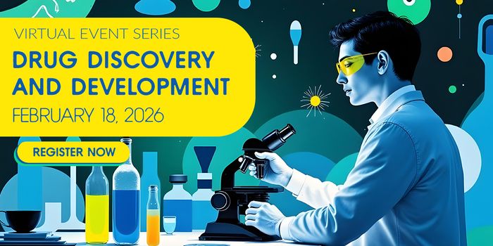
Experience the forefront of pharmaceutical innovation as Labroots and the Drug Discovery and Development planning committee host the 9th Annual Event in the Drug Discovery & Development...
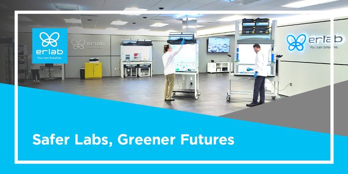
As global sustainability targets and regulatory standards tighten, laboratory environments must evolve to meet both safety and environmental performance expectations. In this thought-provoki...
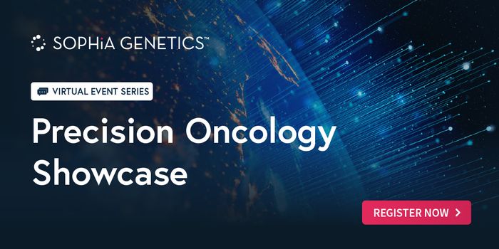
The Precision Oncology Showcase is a webinar series sponsored by SOPHiA GENETICS, set to redefine how precision oncology is practiced worldwide. Taking place throughout 2025, this series wil...

Labroots is thrilled to announce the 14th Annual Event in the Neuroscience Virtual Event Series , streaming live on March 4th, 2026 . Join us as we dive into the captivating world of neurosc...
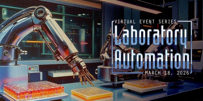
Labroots is delighted to host the 10 th Annual Event in the Laboratory Automation Virtual Event Series , scheduled on March 18th, 2026 ! Dive into the cutting edge of laboratory technology a...
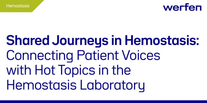
Join a global symposium exploring Hemophilia and Antiphospholipid Syndrome (APS) with patient stories and expert insights for the Hemostasis laboratory....
Agenda Share
-
SEP 26, 2019 1:30 PM PDT
Identification of Novel Biomarkers for Early Diagnosis and Prognosis of Melanoma
-
SEP 26, 2019 1:30 PM PDT
The Visium Spatial Gene Expression Solution: Gene Expression with Spatial Context
-
SEP 26, 2019 12:00 PM PDT
Understanding the Regulation of Membrane Curvature During Mating in Tetrahymena Thermophila
-
SEP 26, 2019 10:30 AM PDT
Differential effects of OCRL1 mutations in Lowe Syndrome cellular phenotypes
Swetha Ramadesikan
Graduate Student in the Aguilar lab at the Department of Biological Sciences, Purdue UniversityBIOGRAPHY -
SEP 26, 2019 10:30 AM PDT
Discover the genes that matter while preserving spatial information: The Visium Gene Expression Solution
-
-
SEP 26, 2019 9:00 AM PDT
Identification of Novel Biomarker Candidates for Early Detection of Melanoma
Margaret Hoang, PhDSenior Scientist, Research & Development, NanoString Te...
Maija Kiuru, PhDAssistant Professor of Clinical Dermatology and Patholo... -
SEP 26, 2019 7:30 AM PDT
Keynote Presentation: Phagocytosis, macropinocytosis and the innate immune response; recent advances
Sergio Grinstein, PhD
Senior Scientist at The Hospital for Sick Children and Professor of Biochemistry, University of Toronto.BIOGRAPHY -
SEP 26, 2019 6:00 AM PDT
Profiling Focal Areas of FFPE Tissue: Spatial Transcriptomics and the Challenge of Low Input Samples
Bruce Seligmann, PhD
Co-Founder and CSO, BioSpyder Technologies, Inc., Professor, University of Arizona College of PharmacyBIOGRAPHY -
SEP 26, 2019 6:00 AM PDT
The reciprocal relationship between cytokine dysregulation and binge alcohol consumption
S. Alex Marshall, PhD
Assistant Professor, Department of Biology, North Carolina Central UniversityBIOGRAPHY


![Bacteria, environment and function: the basis of microb[iom]e annotation](https://d3bkbkx82g74b8.cloudfront.net/eyJidWNrZXQiOiJsYWJyb290cy1pbWFnZXMiLCJrZXkiOiJ3ZWJpbmFyX3Byb2ZpbGVfaW1hZ2VfMjJhYWY2ZTZlM2JjYjA3OWI0MGI2MWUzYmFiYWFiZDFhNDIxODM4NF84NjM1LmpwZyIsImVkaXRzIjp7InRvRm9ybWF0IjoianBnIiwicmVzaXplIjp7IndpZHRoIjoyMDAsImhlaWdodCI6MjAwLCJmaXQiOiJjb3ZlciIsInBvc2l0aW9uIjoiY2VudGVyIiwiYmFja2dyb3VuZCI6IiNmZmYifSwiZmxhdHRlbiI6eyJiYWNrZ3JvdW5kIjoiI2ZmZiJ9fX0=)

- Spatial Omics
-
SEP 26, 2019 6:00 AM PDT
Profiling Focal Areas of FFPE Tissue: Spatial Transcriptomics and the Challenge of Low Input Samples
Bruce Seligmann, PhD
Co-Founder and CSO, BioSpyder Technologies, Inc., Professor, University of Arizona College of PharmacyBIOGRAPHY -
SEP 26, 2019 9:00 AM PDT
Identification of Novel Biomarker Candidates for Early Detection of Melanoma
Margaret Hoang, PhDSenior Scientist, Research & Development, NanoString Te...
Maija Kiuru, PhDAssistant Professor of Clinical Dermatology and Patholo... -
SEP 26, 2019 10:30 AM PDT
Discover the genes that matter while preserving spatial information: The Visium Gene Expression Solution
-
SEP 26, 2019 12:00 PM PDT
Understanding the Regulation of Membrane Curvature During Mating in Tetrahymena Thermophila
- Cell Biology of Genetic Diseases
-
SEP 26, 2019 6:00 AM PDT
The reciprocal relationship between cytokine dysregulation and binge alcohol consumption
S. Alex Marshall, PhD
Assistant Professor, Department of Biology, North Carolina Central UniversityBIOGRAPHY -
SEP 26, 2019 10:30 AM PDT
Differential effects of OCRL1 mutations in Lowe Syndrome cellular phenotypes
Swetha Ramadesikan
Graduate Student in the Aguilar lab at the Department of Biological Sciences, Purdue UniversityBIOGRAPHY - Cell Biology of Diseases
-
SEP 26, 2019 7:30 AM PDT
Keynote Presentation: Phagocytosis, macropinocytosis and the innate immune response; recent advances
Sergio Grinstein, PhD
Senior Scientist at The Hospital for Sick Children and Professor of Biochemistry, University of Toronto.BIOGRAPHY - Microbiome
-
- Spatial Transcriptomics Technology
-
SEP 26, 2019 1:30 PM PDT
Identification of Novel Biomarkers for Early Diagnosis and Prognosis of Melanoma
Speakers Share
-
Sergio Grinstein, PhD
Senior Scientist at The Hospital for Sick Children and Professor of Biochemistry, University of Toronto.
BIOGRAPHY
-
Zachary Bent
Director - Consumables, Product Development, 10x Genomics
BIOGRAPHY
-
Sabrice Guerrier, PhD
Associate Professor of Biology, Millsaps College
BIOGRAPHY
-
Margaret Hoang, PhD
Senior Scientist, Research & Development, NanoString Technologies, Inc.
BIOGRAPHY
-
Maija Kiuru, PhD
Assistant Professor of Clinical Dermatology and Pathology, UC Davis
BIOGRAPHY
-
S. Alex Marshall, PhD
Assistant Professor, Department of Biology, North Carolina Central University
BIOGRAPHY
-
Swetha Ramadesikan
Graduate Student in the Aguilar lab at the Department of Biological Sciences, Purdue University
BIOGRAPHY
-
Nikhil Rao
Sr. Product Manager, Visium Spatial Gene Expression Solution, 10x Genomics
BIOGRAPHY
-
Bruce Seligmann, PhD
Co-Founder and CSO, BioSpyder Technologies, Inc., Professor, University of Arizona College of Pharmacy
BIOGRAPHY
-
Chengsheng Zhu, PhD
Postdoctoral Associate, Rutgers University
BIOGRAPHY


Posters
Share
Program Committee Share
-
Antonio Baines
Dr. Antonio T. Baines is an Associate Professor in the Department of Biology at North Carolina Central University (NCCU) and an adjunct professor in the Department of Pharmacology in the School of Medicine at the University of North Carolina (UNC) Chapel Hill. He earned a ...
See more -
R. Claudio Aguilar, Ph.D.
Dr. Aguilar obtained his PhD degree in Immunochemistry from the School of Pharmacy and Biochemistry, University of Buenos Aires, Argentina. Dr. Aguilar pursued his post-doctoral training at the National institutes of Health in Bethesda, MD in the lab of the well-known cell ...
See more -
Matthew Flegal
Matt entered the research field over 20 years ago as a lab animal technician at the TSI/Mason contract research facility. He has worked at both contract facilities such as TSI and OREAD Biosafety as well in industry at Pharmacia, Pfizer, and Sanofi-Aventis. During that period he ...
See more -
Brian McNally
Brian McNally, PhD is a Vice President at Kx Advisors and is based in Washington, DC. Brian brings his deep expertise in diagnostics and life sciences to create tailored growth strategies for his clients. He specializes in corporate strategy, product development, market sizing ...
See more

Event Series
September 15, 2027 5:30 am Pacific Time
Cell Biology Virtual Event Series 2027

September 16, 2026 5:30 am Pacific Time
Cell Biology Virtual Event Series 2026
September 17, 2025 6:00 am Pacific Time
Cell Biology Virtual Event Series 2025

September 18, 2024 6:00 am Pacific Time
Cell Biology Virtual Event Series 2024
September 20, 2023 5:30 am Pacific Time
Cell Biology Virtual Event Series 2023
September 21, 2022 5:30 am Pacific Time
Cell Biology Virtual Event Series 2022

September 22, 2021 5:30 am Pacific Time
Cell Biology Virtual Event Series 2021
September 23, 2020 5:30 am Pacific Time
Cell Biology Virtual Event Series 2020

September 26, 2018 6:00 am Pacific Time
Cell Biology Virtual Event Series 2018
September 27, 2017 6:00 am Pacific Time
Cell Biology Virtual Event Series 2017
Help
Share
Loading Comments...